Figures & data
Figure 1. Initial appearance of the lesion in preoperative (A) and postoperative conditions (B).
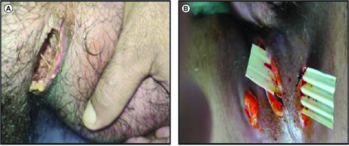
Figure 2. Rapid tumor evolution from September 2022 (A) to December 2023 (B).
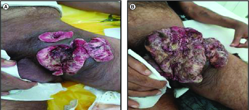
Figure 3. Radiological appearance of the tumor.
(A) MRI: Axial T2-weighted section centered on the pelvic region: large left inguinal mass in T2 hypoposignal (yellow arrow). (B) CT scan of the tumor coronal reconstruction without injection of contrast medium: left inguinal mass spontaneously hypodense (yellow arrow).
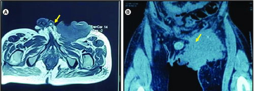
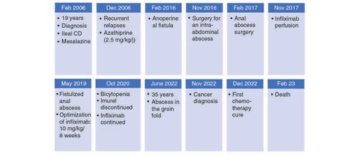
Figure 4. Clinical history of the patient.
CD: Crohn's disease.