
Figures & data
Figure 1. Coronal and axial scan.
(A) Nodular thickening in the transverse and right colon, (B) adrenal nodes (23 × 31 mm) and latero cava lymphadenopathy, (C) thoracic CT scan shows no lymphadenopathy.
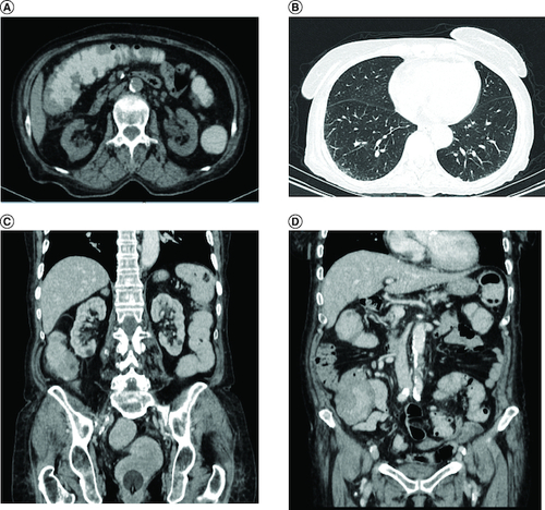
Figure 2. Polypoid mass lesion with ulceration.
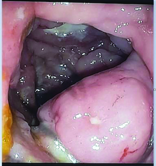
Figure 3. Histological examination.
(A) A diffuse infiltrate of the colon mucosa that didn't invade crypts (HE × 200). (B) The infiltrate was composed of medium to large cells with atypical nuclei and numerous mitoses (HE × 400).
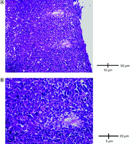
Figure 4. Immunohistological analysis.
(A) Cells were diffusely positive for CD4 (IHC × 200). (B) Cells were diffusely positive for CD8 (IHC × 200).
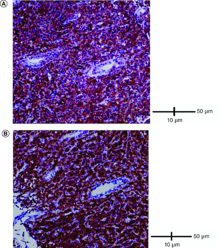
Figure 5. Immunohistological analysis.
(A) Cells were negative for TdT (IHC × 400). (B) Cells were negative for ALK (IHC × 400).
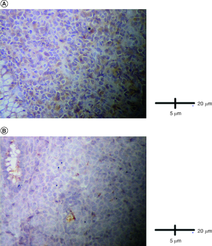
Figure 6. Ulcero-budding gum tumor with repression of the fixed prosthesis.
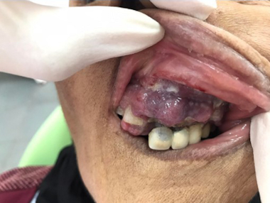
Table 1. Case reports of intestinal T-cell lymphomas not otherwise specified.