Figures & data
Figure 1. In a 400-fold magnified photo of human umbilical cord mesenchymal stem cells under an inverted microscope.
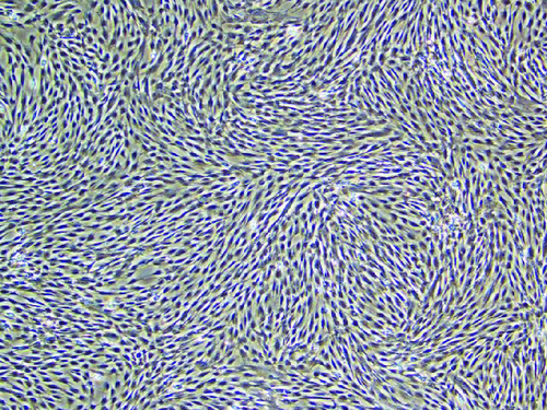
Figure 2. Positive indicators CD73+, CD90+ and CD105+ are all greater than 95.0%, while the negative indicators HLA-DR+, CD11+ and CD45+ are all less than 2%, meeting the standard.
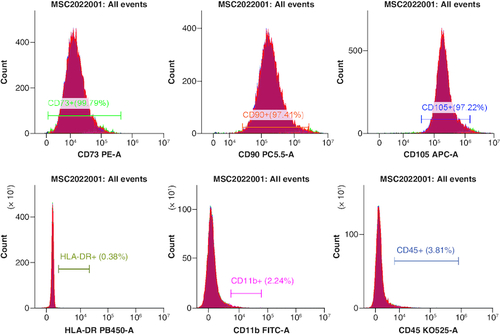
Figure 3. Electron microscopy.
(A) Small porcine intestinal submucosa. (B) Human umbilical cord mesenchymal stem cells combined with small porcine intestinal submucosa (scale bar = 100 μm).
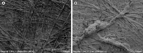
Figure 4. The effect of human umbilical cord mesenchymal stem cells combined with porcine small intestinal submucosa on wound healing.
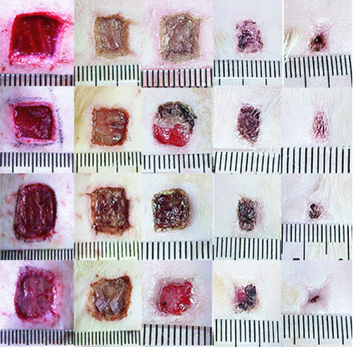
Table 1. Wound area after treatment (cm2), (±s, blank control group n = 5, small porcine intestinal submucosa group, human umbilical cord mesenchymal stem cell group, and human umbilical cord mesenchymal stem cells combined with small porcine intestinal submucosa group. n = 10).
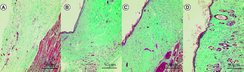
Figure 5. Effect of human umbilical cord mesenchymal stem cells combined with small porcine intestinal submucosa on inflammatory cells (H&E staining ×200).
(A) Blank control group. (B) Small porcine intestinal submucosa group. (C) Human umbilical cord mesenchymal stem cell group. (D) Human umbilical cord mesenchymal stem cells combined with small porcine intestinal submucosa group (scale bar = 50 μm).
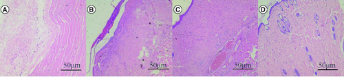
Figure 6. Effect of human umbilical cord mesenchymal stem cells combined with small porcine intestinal submucosa on wound microvessel density (CD31, ×200 magnification).
(A) Blank control group. (B) Small porcine intestinal submucosa group. (C) Human umbilical cord mesenchymal stem cell group. (D) Human umbilical cord mesenchymal stem cells combined with small porcine intestinal submucosa group (scale bar = 50 μm).
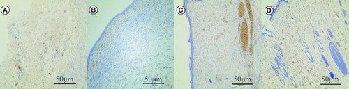
Figure 7. Effect of human umbilical cord mesenchymal stem cells combined with small porcine intestinal submucosa on wound collagen fibers (Masson staining, ×200).
(A) Blank control group. (B) Small porcine intestinal submucosa group. (C) Human umbilical cord mesenchymal stem cell group. (D) Human umbilical cord mesenchymal stem cells combined with small porcine intestinal submucosa group.