Figures & data
Figure 1. Whole body positron emission tomography-computed tomography before treatment.
(A–D) Whole body PET-CT showed that enlarged hypermetabolic mediastinum lymph nodes(SUVmax:9.2). (E–H) Whole body PET-CT showed that a highly metabolic irregular mass in right kidney with heterogeneous density (120 mm*108 mm, SUVmax:15.2) and the border demarcation between the masses and the liver were indistinct.
PET-CT: Positron emission tomography-computed tomography.
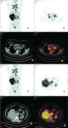
Figure 2. Radiological evaluation before and after treatment.
(A) Abdominal MRI revealed irregular mass in right kidney with heterogeneous density and the soft tissue density shadows was seen in the inferior vena cava and right renal vein on 3 October 2022. (B) CT-SCAN revealed multiple enlarged mediastinum lymph node metastasis. (C) Abdominal MRI revealed irregular mass in right kidney with heterogeneous density significant reduction after two cycles of treatment. (D) CT-SCAN revealed significant mediastinum lymph node almost disappeared after two cycles treatment. (E) Abdominal MRI revealed irregular mass in right kidney with heterogeneous density continued reduction after four cycles of treatment. (F) CT-SCAN revealed significant mediastinum lymph node almost disappeared after four cycles of treatment.
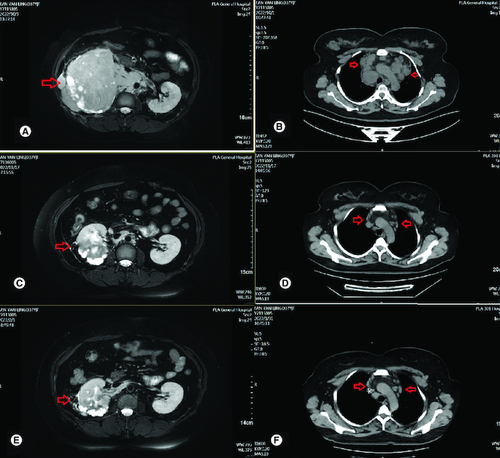
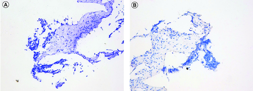
Figure 3. Staining images of PD-L1 expression (HE and IHC).
(A) HE staining images of PD-L1 expression. (B) IHC staining images of PD-L1 expression.
HE: Hematoxylin-eosin staining; IHC: Immunohistochemistry.