Figures & data
Table 1. Clinical and endoscopic characteristics of patients.
Figure 1. Endoscopic aspects.
(A) Slightly elevated whitish–yellow plaques. (B) Confluent plaques creating a pseudomembranous aspect, sometimes surrounded by erythematous colonic mucosa (blue arrows). (C) Formation of plaques rapidly after the appearance of whitish foamy fluid.
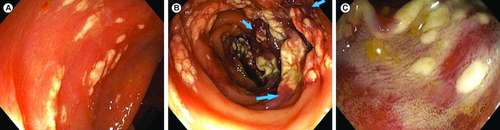
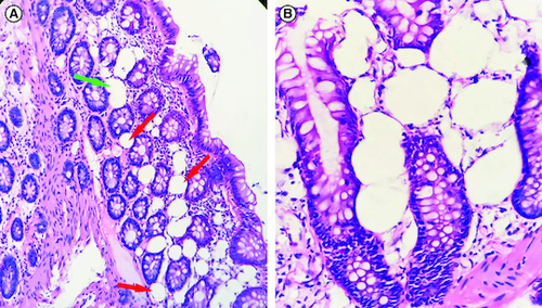
Figure 2. Histological examination.
(A) Numerous optically empty vacuoles of variable sizes (red arrows) in the lamina propria and the muscularis mucosa, sometimes confluent (green arrow), intermingled with inflammatory cells (hematoxylin-eosin 200×). (B) The optically empty vacuoles repress and distort the cryptic contours (hematoxylin-eosin 400×).