Figures & data
Figure 1. FT-IR spectrum of isolated compound which corresponds with molecular structure of cyanidin.
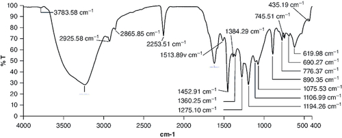
Figure 2. Body weight changes during 14 days of acute oral toxicity test.
The data (n = 3) is shown as mean ± standard deviation. Two-way analysis of variance was used for the comparative analyses, and Tukey's test was used for the pairwise post-hoc analysis. A p-value of less than 0.05 was used as the threshold for statistical significance.
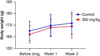
Figure 3. Body weight changes during 28 days of subacute oral toxicity test.
The data is shown as mean ± standard deviation (n = 5). Two-way analysis of variance was used for the comparative analyses, and Tukey's test was used for the pairwise post-hoc analysis. A p-value of less than 0.05 was used as the threshold for statistical significance. ‘M’ represents male group and ‘F’ represents female group.
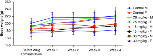
Table 1. Results of acute toxicity test.
Table 2. Weight of all major organs measured immediately at the end of the study.
Table 3. Impact of cyanidin on experimental and control groups on hematological parameters.
Table 4. Impact of cyanidin on experimental and control groups on liver function.
Table 5. Effect of cyanidin on renal function of control and experimental groups.
Table 6. Effect of cyanidin on serum cholesterol and blood glucose levels of control and experimental groups.
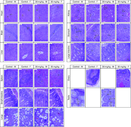
Figure 4. Presents photomicrographs showcasing significant organs from both genders in the high-dose and control groups.
Staining was performed using hematoxylin and eosin, with a scale bar set at 800 μm. Morphology of all major organs of treated groups represents normal architecture comparable to that of control groups.
Heart – MF: Myocardial fiber; N: Nucleus; Brain – GL: Granular layer; ML: Molecular layer; PN: Pyramidal neuron; Liver – CV: Central vein; S: Sinusoid; Kidney – PCT: Proximal convoluted tubule; RC: Renal corpuscle; RT: Renal tubule; Stomach – Ccell: Chief cell; GP: Gastric pit; LP: Lamina propria; Pcell: Parietal cell; Large intestine – IG: Intestinal gland; GC: Goblet cell; L: Lumen; M: Mucosa; SM: Submucosa; Spleen – CA: Central arteriole; RP: Red pulp; WP: White pulp; Small intestine – C: Crypt; SM: Submucosa; V: Villi; Lungs – B: Bronchiole; BV: Blood vessel; PA: Pulmonary alveoli; Ovary – G: Granulosa cell; GF: Graafian follicle; OC: Oocyte; Testis – SP: Spermatocyte; ST: Seminiferous tubule; SZ: Spermatozoa.