Figures & data
Figure 1 Cell proliferation inhibition profiles of ChalcEA in T47D cell lines.
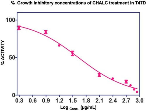
Figure 2 (A) Pharmacophore features of 4-OHT (yellow-colored sticks) in hERα (3ERT) and (B) in comparison with the docking pose of ChalcEA (cyan-colored sticks).
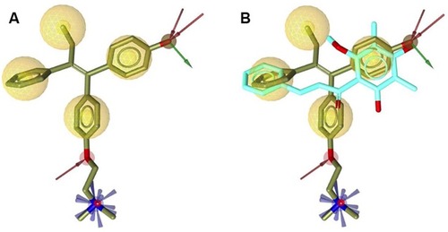
Figure 3 Re-docking of co-crystallized 4-OHT into the ligand-binding site of hERα using AutoDock 4.2 (crystal and docking poses are colored in grey and orange, respectively).
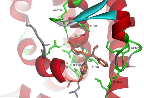
Figure 4 The superimposition between the docking poses of ChalcEA and 4-OHT (blue and orange-colored sticks, respectively) inside the ligand-binding site of hERα.
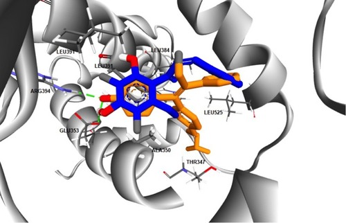
Figure 5 RMSD profile of 3ERT-ChalcEA and 3ERT-4OHT during 20 ns of MD simulation.
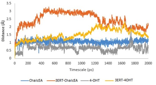
Figure 6 RMSF profile of 3ERT-ChalcEA and 3ERT-4OHT systems throughout 20 ns of MD simulation.
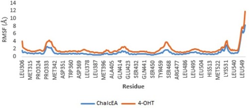
Figure 7 The plot of dihedral analysis of His524’s side-chain rotation during 20 ns of simulation.
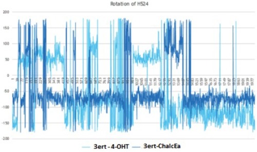
Table 1 Van Der Waals (VDW) Analysis Of The ChalcEA And 4-OHT System In Both Antagonist Receptor Forms Throughout 20 ns Of MD Simulation
Figure 8 ROC curve for validation of the pharmacophore model by using the 625-compound active set and the 20,722-compound decoy set.
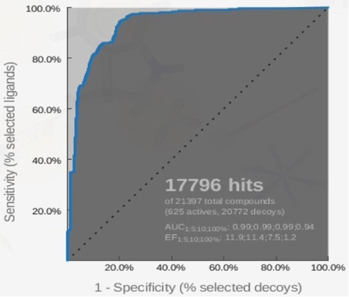
Table 2 IC50 Of Chalcone Derivatives Against T47D Human Breast Cancer Cell Lines
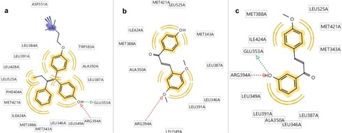
Figure 9 Molecular docking results of chalcone derivatives against human estrogen receptor alpha (HERα). (A) Hydroxyl group of 4-OGT formed hydrogen bonds with GLU353 and Arg394 of HERα, (B) while CL4 was play role as hydrogen bond acceptor against Arg394, and (C) 4-OMe also formed hydrogen bonds with GLU353 and Arg394 of HERα.