Figures & data
Figure 1 Structure variability within a protein family.
Notes: Structures of acetylcholinesterase (1acg, 1ax9, 1dx6, 1hbj, 1qon, 1vot, and 2ace) crystallized with different active-site ligands.
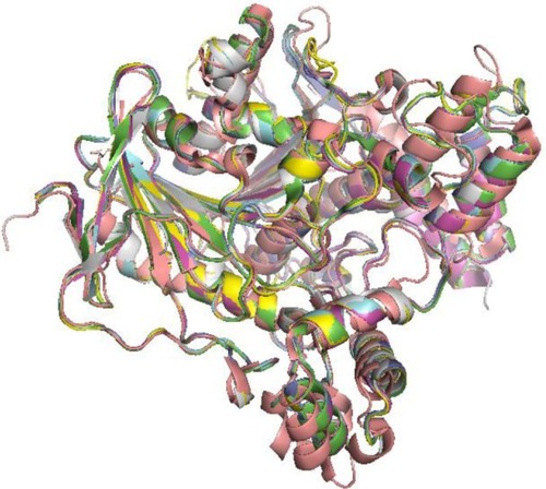
Figure 2 Experimental ensembles.
Notes: (A) Superimposition of experimental structures of protein kinases. (B) NMR derived ensemble of calcium-binding protein (PDB code: 1A03).
Abbreviations: NMR, nuclear magnetic resonance; PDB, protein data bank.
Abbreviations: NMR, nuclear magnetic resonance; PDB, protein data bank.
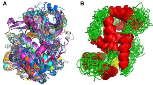
Figure 3 Molecular dynamics basic algorithm.
Notes: The simulation output, the trajectory, is an ordered list of 3N atom coordinates for each simulation time (or snapshot).
Abbreviations: Epot, potential energy; t, simulation time; dt, iteration time; For each spatial coordinate of the N simulated atoms (i): x, atom coordinate; F, forces component; a, acceleration; m, atom mass; v, velocity.
Abbreviations: Epot, potential energy; t, simulation time; dt, iteration time; For each spatial coordinate of the N simulated atoms (i): x, atom coordinate; F, forces component; a, acceleration; m, atom mass; v, velocity.
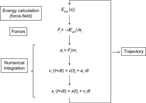
Figure 4 R-T transition on Bacillus stearothermophilus lactate dehydrogenase after 50 ns simulation in explicit solvent.
Notes: Inset: detail of conformational shift of active site substrate-binding Arg 169 side chain. Simulation was initiated from a dimeric model of the protein (1ldn), and allowed to evolve without restrains. Protein system was prepared using MDWeb,Citation91 and simulated using GROMACS,Citation148 at 298 K of temperature, in explicit solvent and periodic boundary conditions (truncated octahedron box). Conformational shift is indicated as R-T shift in the main figure and with an arrow in the inset.
Abbreviation: R-T, relaxed and tense states (as defined by Monod’s model).
Abbreviation: R-T, relaxed and tense states (as defined by Monod’s model).
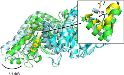
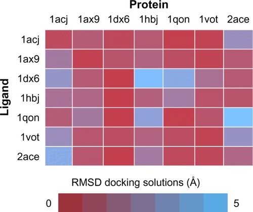
Figure 5 Cross-docking experiment with selected acetylcholinesterase structures from PDB.
Notes: Performance of all possible combinations of rigid docking experiments done in standard conditions, using a series of seven acetylcholinesterase ligands extracted from left column PDB entries onto the same empty protein structures (upper row). Color code indicates RMSD between the best docked solution and the reference PDB.
Abbreviations: PDB, protein data bank; RMSD, root-mean-square deviation.
Abbreviations: PDB, protein data bank; RMSD, root-mean-square deviation.