Figures & data
Figure 1 Schematics of the major bioprinting mechanisms.
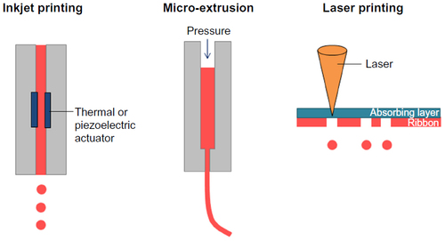
Table 1 Comparison of major bioprinting techniques
Table 2 List of commonly used biomaterials for bioprinting
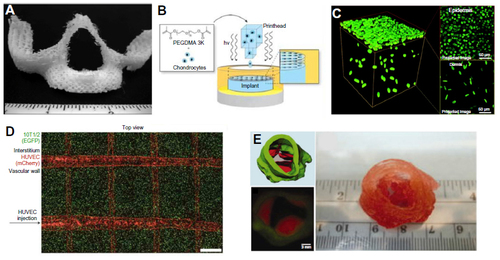
Figure 2 Examples of the bioprinted tissues.
Notes: (A) 3D-printed, porous PCL scaffolds of maxilla at 40% infill density. Copyright © 2014. John Wiley and Sons. Reproduced from Temple JP, Hutton DL, Hung BP, et al. Engineering anatomically shaped vascularized bone grafts with hASCs and 3D-printed PCL scaffolds. J Biomed Mater Res A. 2014;102(12):4317–4325.Citation43 (B) Schematic of bioprinting cell-laden constructs for cartilage tissue engineering with simultaneous photopolymerization process. Copyright © 2012. John Wiley and Sons. Reproduced from Cui X, Breitenkamp K, Lotz M, D’Lima D. Synergistic action of fibroblast growth factor-2 and transforming growth factor-beta1 enhances bioprinted human neocartilage formation. Biotechnol Bioeng. 2012; 109(9):2357–2368.Citation47 (C) 3D-printed skin tissue with keratinocyte-populated epidermal layer and fibroblast-populated dermal layer. Scale bar, 500 μm. Copyright © 2014, Mary Ann Liebert, Inc. Reproduced from Lee V, Singh G, Trasatti JP, et al. Design and fabrication of human skin by three-dimensional bioprinting. Tissue Eng Part C Methods. 2014;20(6):473–484.Citation53 (D) 3D vascular network (red) with 10T1/2 cells (green) in the interstitial space. Scale bar, 1 mm. Reprinted by permission from Macmillan Publishers Ltd: Nature Materials. Miller JS, Stevens KR, Yang MT, et al. Rapid casting of patterned vascular networks for perfusable engineered three-dimensional tissues. Nat Mater. 2012;11(9):768–774, copyright © 2012.Citation61 (E) Bioprinting of aortic valve conduit with dual cell types for valve root (green, smooth muscle cells) and valve leaflets (red, valve leaflet interstitial cells). Copyright © 2013. John Wiley and Sons. Reproduced from Duan B, Hockaday LA, Kang KH, Butcher JT. 3D bioprinting of heterogeneous aortic valve conduits with alginate/gelatin hydrogels. J Biomed Mater Res A. 2013;101(5):1255–1264.Citation75
Abbreviations: HUVEC, human umbilical vein endothelial cell; PCL, polycaprolactone; PEGDMA, polyethylene glycol dimethacrylate; 3D, three-dimensional; EGFP, enhanced green fluorescent protein.
Abbreviations: HUVEC, human umbilical vein endothelial cell; PCL, polycaprolactone; PEGDMA, polyethylene glycol dimethacrylate; 3D, three-dimensional; EGFP, enhanced green fluorescent protein.