Figures & data
Figure 1 Breast cancer cells functionally express the CB2 receptor.
Notes: (A) Western blot of cell lysates from mouse and human breast cancer cell lines showing expression of the CB2 receptor (top) and loading control α-tubulin (bottom). (B) In vitro cAMP ELISA performed on 4T1 cells after 30-minute incubation with phosphodiesterase inhibitor IBMX and 30-minute stimulation with either positive control Fsk, control Veh, or JWH-015 (range 10 pM to 10 µM) (*P<0.05). (C) Sulforhodamine B-cell viability assays after shRNA DNA lentiviral knockdown of CB2 receptor. Cell viability 60 hours after a 12-hour incubation period with indicated concentrations of anti-CB2 shRNA shown (*P<0.05). (D) Western blot showing degree of CB2 knockdown after incubation with shRNA DNA lentiviral particles.
Abbreviations: CB2, cannabinoid receptor 2; GAPDH, glyceraldehyde phosphate dehydrogenase; cAMP, cyclic adenosine monophosphate; shRNA, small hairpin RNA; ELISA, enzyme-linked immunosorbent assay; IBMX, 3-isobutyl-1-methylxanthine; Fsk, forskolin; Veh, vehicle.
Abbreviations: CB2, cannabinoid receptor 2; GAPDH, glyceraldehyde phosphate dehydrogenase; cAMP, cyclic adenosine monophosphate; shRNA, small hairpin RNA; ELISA, enzyme-linked immunosorbent assay; IBMX, 3-isobutyl-1-methylxanthine; Fsk, forskolin; Veh, vehicle.
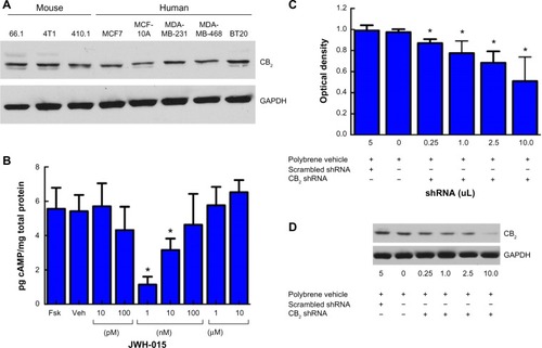
Figure 2 In vivo administration of JWH-015 attenuates primary mammary tumor growth.
Notes: (A) Weekly in vivo bioluminescence imaging representative of tumor progression in immunocompetent mice receiving either vehicle (top) or JWH-015 (6 mg/kg) for 21 days beginning 7 days after tumor inoculation (1×10⁶ 4T1 cells via orthotopic injection into the c-9 mammary fat pad) in the 4T1-Luc murine mammary carcinoma model. (B) Day 28 mass of resected primary tumors from vehicle- (left) or JWH-015-treated mice (*P<0.05). (C) Mice were evaluated for metastasis via luminometer analysis of cell lysates isolated from common metastatic sites on day 28.
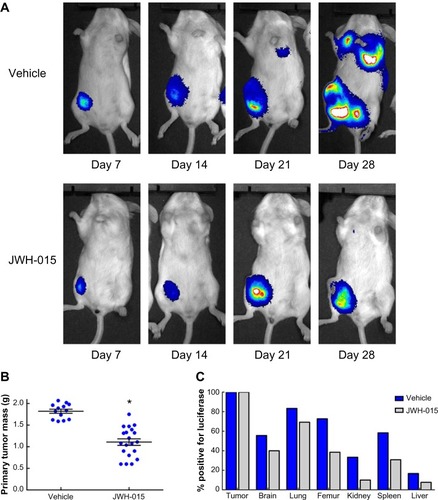
Figure 3 CB2 inverse agonist SR144528 does not attenuate cell viability reduction induced by JWH-015 in vitro.
Notes: Sulforhodamine B assays for cell viability were performed on murine 4T1 (A, B) and human MCF7 (C, D) breast cancer cells after administration of either vehicle, JWH-015 alone, SR144528 alone, or JWH-015 and SR144528 in combination. Cell viability in response to 48-hour incubation with JWH-015 is shown (A, C; *P<0.05). Dose–response curves in response to all treatment groups are depicted in panels (B) and (D) for comparison.
Abbreviation: CB2, cannabinoid receptor 2.
Abbreviation: CB2, cannabinoid receptor 2.
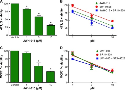
Figure 4 In vitro administration of JWH-015 induces apoptosis of breast cancer cells independent of cell cycle.
Notes: (A) 40× magnification. Ki67 imaging studies were performed on 4T1 cells in response to vehicle (upper panels), 1 µM JWH-015 (middle panels) or 10 µM JWH-015 (lower panels). DAPI nuclear staining (left) is consistent with Ki67 staining (right) in all treatment conditions. Early caspases 3 and 7 were evaluated via ELISA in 4T1 cells (B) and MCF7 cells (which express caspase 7 but not caspase 3); (C) after 24 hours of exposure to vehicle or 1, 3, or 10 µM JWH-015. Samples were normalized to caspase levels in untreated control samples (*P<0.05).
Abbreviations: DAPI, 4′,6-diamidino-2-phenylindole; ELISA, enzyme-linked immunosorbent assay.
Abbreviations: DAPI, 4′,6-diamidino-2-phenylindole; ELISA, enzyme-linked immunosorbent assay.
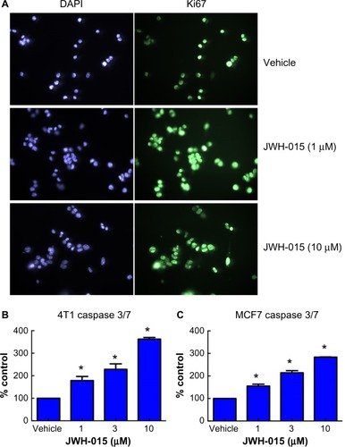
Figure 5 JWH-015 induction of calcium flux and modulation of MAPK/ERK phosphorylation is calcium dependent.
Notes: Fura 2-AM signal was recorded every 30 seconds for a period of 60 minutes in 4T1 cells treated with vehicle (A) or 10 µM JWH-015 (C). Graphs are depicted as ratio of recordings at 340 over 380 nm vs time. Quantification of signal (B, D) shows an initial inhibition of calcium efflux followed by a marked increase in efflux 60 minutes postadministration of JWH-015 (*P<0.05). Western blot analysis of phospho-ERK1/2 (E) demonstrates JWH-015 inhibition of the MAPK/ERK pathway that is attenuated when culture media is replaced with calcium-depleted media 20 minutes prior to administration of 10 µM JWH-015. ImageJ quantification of the blots normalized to α-tubulin loading control is depicted in panel (F) (*P<0.05 in Opti-MEM cultured cells and **P<0.05 in calcium-depleted cultures).
Abbreviations: Opti-MEM, Opti-minimal essential medium; Fura 2-AM, Fura 2-acetoxymethyl.
Abbreviations: Opti-MEM, Opti-minimal essential medium; Fura 2-AM, Fura 2-acetoxymethyl.
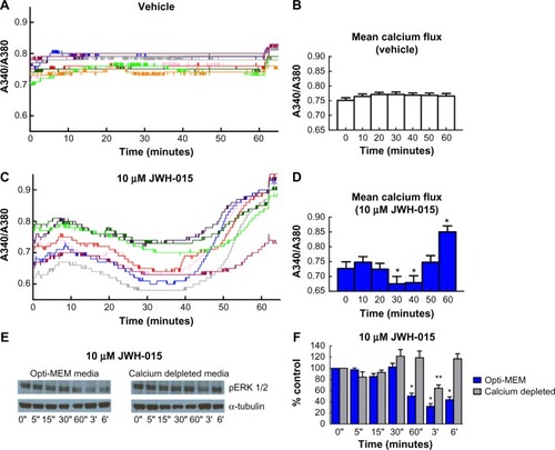
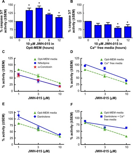
Figure 6 Inhibition of calcium flux attenuates JWH-015-induced apoptosis in 4T1 cells.
Notes: Timeline analysis of caspase 3/7 protein expression in 4T1 cells in response to 10 µM JWH-015 in normal media (A) or calcium-depleted media (B) demonstrates ablation of JWH-015 effects when performed in calcium-depleted media (*P<0.05). Relevance of extracellular calcium flux vs intracellular calcium flux was evaluated pharmacologically by performing SRB assays on 4T1 cells in response to JWH-015 in the presence of L-type calcium-channel blocker nifedipine, N/P/Q-type calcium-channel blocker ω-Conotoxin MIIVC (C), normal vs calcium-depleted media (D), intracellular ryanodine receptor antagonist dantrolene (E), and calcium depletion concurrent with dantrolene (F).
Abbreviations: SEM, standard error of mean; Opti-MEM, Opti-minimal essential medium; SRB, sulforhodamine B.
Abbreviations: SEM, standard error of mean; Opti-MEM, Opti-minimal essential medium; SRB, sulforhodamine B.