Figures & data
Table 1 Antibodies used
Figure 1 Confocal microscopy images of triple immunofluorescence-labeled histologically normal breast tissue.
Notes: (A)–(E) Images of single optical sections (0.6 µm thick) from a stromal area with a duct visible in the upper left part of the field (* in lumen), demonstrating immunofluorescence multilabeling and DAPI (nuclear stain) in separate channels. (A) ALDH1, green, indicates the different shaped ALDH1+ cell types in stroma (round/oval cells [short arrows] and spindle-shaped/polygonal cells [long arrows]). (B) CD24, red, indicates the scarcity of CD24+ cells. (C) CD44, white, indicates the relatively large number of CD44+ cells, yet many ALDH1+ CD44− spindle-shaped cells. (D) DAPI, blue. (E) All four channels merged indicate that many ALDH1+ cells are also CD44+ but not CD24+. Arrows indicate cell types as in (A). (F) Confocal image (merged channels are color coded as in A–E) from a different tissue area showing multilabeled breast tissue containing the stromal cell types as described in A–E, as well as ductules and numerous adjacent ALDH1+ CD24− cells with weak membranous CD44 reactivity. Scale bars = 10 µm.
Abbreviation: DAPI, 4′,6-diamidino-2-phenylindole.
Abbreviation: DAPI, 4′,6-diamidino-2-phenylindole.
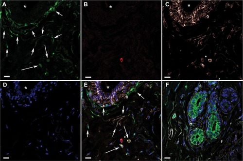
Table 2 Genetic and hormonal characteristics, and cancer status of the patients
Figure 2 ALDH1+ cells in TDLUs.
Notes: Fluorescence microscope images of a triple-labeled TDLU from benign breast tissue with the following color coding: (A) DAPI nuclear staining (blue); (B) ALDH1 (green); (C) CD44 (white); (D) CD24 (red). (E) A digitally composed image showing all channels merged. Most of the cells located basally in ductules are ALDH− CD44+, whereas some adluminal cells are ALDH1+ CD44+ and none are CD24+. In stroma, elongated ALDH1+ cells are seen between the ductules, and many such cells are present in the junction between TDLU stroma and generic connective tissue of the breast (arrows). Scale bars = 20 µm.
Abbreviations: DAPI, 4′,6-diamidino-2-phenylindole; TDLUs, terminal duct-lobular units.
Abbreviations: DAPI, 4′,6-diamidino-2-phenylindole; TDLUs, terminal duct-lobular units.
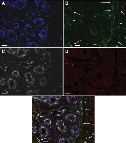
Figure 3 ALDH1+ spindle-shaped/polygonal cells in TDLU stroma (arrows).
Notes: DAPI nuclear staining (blue), ALDH1 (green), CD44 (white), and CD24 (red). Scale bar = 20 µm.
Abbreviations: DAPI, 4′,6-diamidino-2-phenylindole; TDLU, terminal duct-lobular unit.
Abbreviations: DAPI, 4′,6-diamidino-2-phenylindole; TDLU, terminal duct-lobular unit.
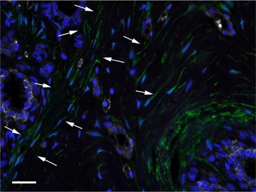
Figure 4 ALDH1+ round/oval-shaped cells in TDLU stroma.
Notes: DAPI nuclear staining (blue) and labeling of ALDH1 (green), CD44 (white), and CD24 (red). (A) A single-channel image depicting ALDH1+ round/oval cells (arrows). (B) A composite image with all channels demonstrating that the ALDH1+ round/oval cells are positive for CD44 but not for CD24 (arrows). Scale bars = 20 µm.
Abbreviations: DAPI, 4′,6-diamidino-2-phenylindole; TDLU, terminal duct-lobular unit.
Abbreviations: DAPI, 4′,6-diamidino-2-phenylindole; TDLU, terminal duct-lobular unit.
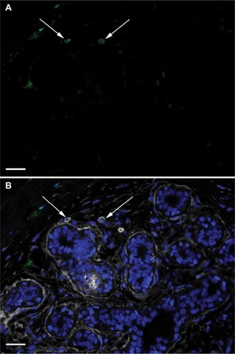
Figure 5 CD24+ cells in TDLU stroma.
Notes: DAPI nuclear staining (blue) and labeling of ALDH1 (green), CD44 (white), and CD24 (red). The image depicts a CD24+ cell (arrow) exhibiting no CD44 or ALDH1 positivity. Scale bar = 20 µm.
Abbreviations: DAPI, 4′,6-diamidino-2-phenylindole; TDLU, terminal duct-lobular unit.
Abbreviations: DAPI, 4′,6-diamidino-2-phenylindole; TDLU, terminal duct-lobular unit.
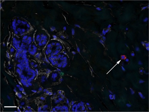
Table 3 Immunohistological results for the patient groups
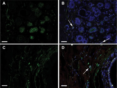
Figure 6 Round/oval cells according to family history.
Notes: DAPI nuclear staining (blue) and labeling of ALDH1 (green), CD44 (white), and CD24 (red). (A)–(B) Images showing the absence of ALDH1+ CD44+ CD24− round/oval cells in a TDLU from a woman with a family history of breast cancer. Arrows indicate ALDH1− CD44+ CD24− cells. (C)–(D) Images illustrate an ALDH1+ CD44+ CD24− round/oval-shaped cell (arrow) in a TDLU from a woman with no family history of breast cancer. (A) and (C) show only the ALDH1+ channel, and (B) and (D) show all four channels merged. Scale bars = 20 µm.
Abbreviations: DAPI, 4′,6-diamidino-2-phenylindole; TDLU, terminal duct-lobular unit.
Abbreviations: DAPI, 4′,6-diamidino-2-phenylindole; TDLU, terminal duct-lobular unit.