Figures & data
Figure 1 The picture shows the lesion characterized by a big morula and many pustules with a frail roof within hemorrhagic fluid, outlined by an edematous circular edge.
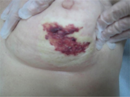
Figure 2 Two months after initial presentation, the involved area became pale, avascular, and covered by a transparent epithelial coat surrounded by multiple frail and thin telangiectasias with retraction of the skin in the inferior quadrants of the breast.
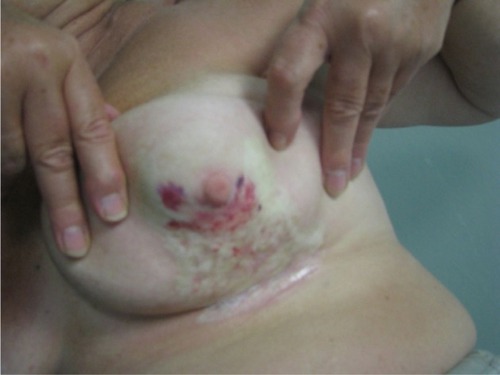
Figure 3 The picture shows a severely retracted and distorted breast with edema of the subcutaneous tissues as well as induration in the central part over the nipple area with peripheral telangiectasias.
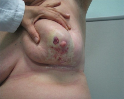
Figure 4 Magnetic resonance images of the breast show a contrast enhancement of the dermal-epidermal surface with thickening of the skin contour. (A) Is in the caudal part where there is oedema, (B) is in the middle part of the lesion where there are more vessels.
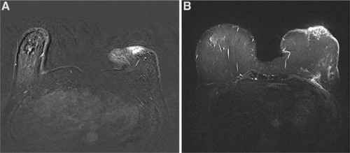
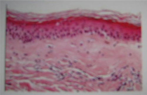
Figure 5 Patch biopsy of the involved area with findings of chronic inflammatory disease evidenced as panniculitis and chronic radiodermatitis.