Figures & data
Figure 1 Transcription and subcellular distribution of TRPV1 in MDA-MB-231 and MCF-7 cell lines.
Notes: (A) TRPV1 mRNA expression in MDA-MB-231 and MCF-7 by qPCR did not reveal any statistical difference. (B) Different abundance and distribution of TRPV1 in MDA-MB-231 and MCF-7. Image magnification: images was obtained in 100· (optical). After analysis process, digital magnification was 1.25·. (C) Colocalization analysis results. In MDA-MB-231, most of TRPV1 protein followed the same subcellular distribution as the ER, whereas the classical pattern in MCF-7 did not colocalize with the ER/Golgi mark. (D) MDA-MB-231 and MCF-7 exhibited different expressions of ERs. In MDA-MB-231, qPCR experiments showed expression of ERβ only, whereas MCF-7 expressed both ERα and ERβ (P<0.05). (E) The transcription of TRPV1 in MCF-7 was induced by estradiol and blocked by the antagonist of ER ICI 182780. *p>0.05. (F) In MCF-7, TRPV1 did not present significant colocalization with any of the masks automatically generated, ie, plasma membrane, cytosol, and nucleus. This pattern can be classified as classical. On the other hand, MDA-MB-231 colocalizes more proteins in the cytosol mask, this pattern being classified as nonclassical.
Abbreviation: qPCR, quantitative PCR.
Abbreviation: qPCR, quantitative PCR.
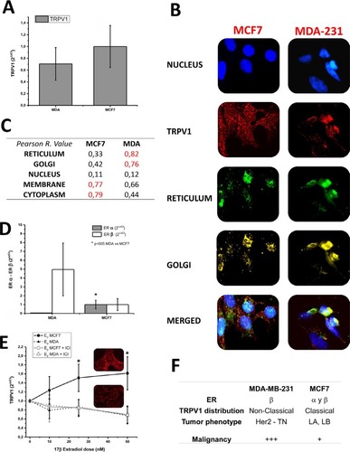
Table 1 Clinical parameters according to TRPV1 expression categories
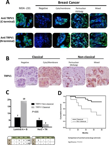
Figure 2 Nonclassical pattern of TRPV1 identifies higher malignancy breast carcinomas.
Notes: (A) The immunofluorescence of C-TRPV1 and N-TRPV1 antibodies confirmed the expression of TRPV1 in breast cancer, with a diffuse expression pattern. (B) The immunohistochemical detection of TRPV1 shows a classical pattern at the plasma membrane and cytosol, and a nonclassical pattern with aggregates of TRPV1 at the ER/Golgi and/or a relatively diffuse distribution of the channel at the surrounding cytosol. (C) The classical TRPV1 pattern was more frequent in lower malignancy St. Gallen subtypes luminal A + luminal B compared to the most aggressive subtypes Her 2 + triple negative. (D) Survival curves according to the pattern of TRPV1 subcellular distribution.