Figures & data
Figure 1 Search strategy.
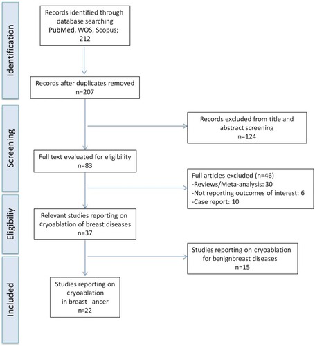
Figure 2 Cryoablation of invasive breast carcinoma in a 74-year-old woman presenting with distant metastases to the bones (Stage IV) at diagnosis. Axial (A) and coronal (B) contrast-enhanced CT scan showing a 4 cm mass in the left breast. (C–D). Contrast-enhanced CT scan 2 months after cryoablation showing tumor size reduction and absence of contrast enhancement.
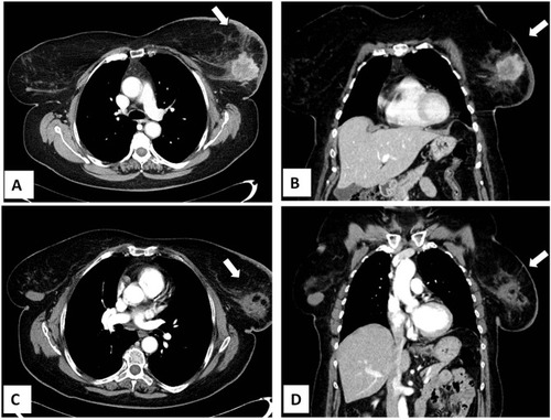
Figure 3 Same patient as shown in . She developed a local recurrence after 12 months from the cryoablation procedure that was treated with redo cryoablation. Axial (A) and coronal (B) contrast-enhanced CT scan images showing a 2 cm nodule with enhancement in the previous ablation zone. (C) Second cryoablation with insertion of two cryo-probes. (D) Complete tumor ablation with no contrast-enhancement at the end of the procedure.
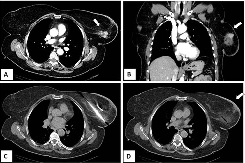
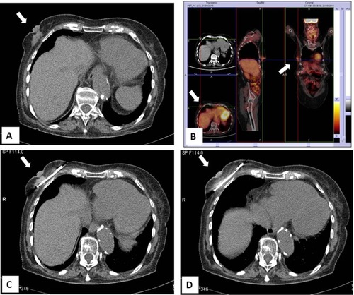
Figure 4 CT-guided cryoablation of large invasive carcinoma of the right breast in 83-year-old woman not suitable for surgery due to co-morbidities. (A) Large tumor mass in the right breast with nipple retraction. (B) PET-FDG/CT imaging showing an abnormal area of18F-FDG uptake in the right breast. (C) CT-guided placement of the cryoprobe. (D) Complete ablation of the tumor during the freezing process.