Figures & data
Figure 1 A 51-year-old female presented with hard right breast after suffering from right breast trauma 2 months previously. (A and B) Craniocaudal and mediolateral mammogram of the right breast revealed a dense breast with architectural distortion noted at upper outer quadrant (more evident on craniocaudal view). (C) Ultrasound revealed an ill-defined irregular hypoechoic mass with posterior acoustic shadowing at 9 o’clock of the right breast.
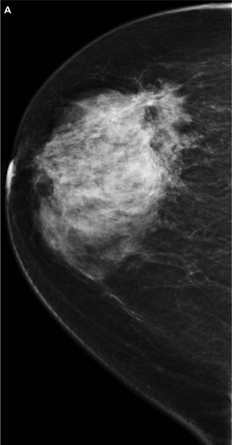
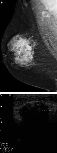
Figure 2 A 48-year-old female patient presented with palpable right axillary lymph node. (A and B) Craniocaudal and mediolateral mammogram revealed asymmetric breast density with no definite masses. (C and D) Ultrasound revealed three small hypoechoic masses at 1–11 o’clock of the right breast (only two are displayed here).
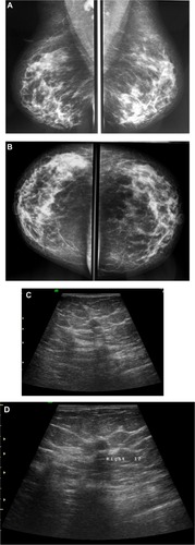
Figure 3 A 58-year-old female with palpable mass in the left breast. (A and B) CC and ML mammogram films revealed ill-defined dense opacity with spiculated margins surrounded by architectural distortion at the lower central area of the breast, which looks to be attached to the pectoralis muscle posteriorly. (C) On ultrasound, ill-defined, spiculated, hypoechoic, solid mass with posterior shadowing measuring 16 × 13 mm is seen at 5 o’clock of the breast.
Abbreviations: CC, craniocaudal; ML, mediolateral.
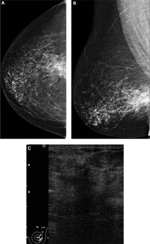
Figure 4 A 72-year-old female with left breast mass. (A and B) Craniocaudal and mediolateral mammogram of left breast revealed an irregular spiculated, dense mass (35 × 25 mm) in the upper outer quadrant close to the pectoral muscle. Single benign macrocalcification is noted in the central area. (C) Ultrasound of the left breast revealed a spiculated, hypoechoic, solid mass (30 × 23 mm) with strong acoustic shadow at 3–4 o’clock.
Abbreviations: CC, craniocaudal; ML, mediolateral.
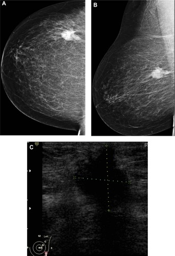
Figure 5 A 48-year-old female patient presented with hard right breast mass of 2 months duration. (A–C) CC, ML, and magnified mammogram views of the right breast revealed an irregular spiculated dense mass at the lower outer quadrant of the right breast. A magnified view shows clusters of pleomorphic microcalcifications within the lesion (arrows). Four foci of calcifications are seen away from the lesion. (D) Ultrasound revealed an ill-defined mass of heterogeneous echopattern at 8 o’clock of left breast.
Abbreviations: CC, craniocaudal; ML, mediolateral.
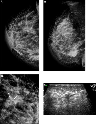
Table 1 Mammographic findings of 32 cases of invasive lobular carcinoma
Table 2 Sonographic findings in 32 cases of invasive lobular carcinoma