Figures & data
Table 1 Comparative Analysis of Age and Mass Size Between PIMPC and NIDC
Table 2 Comparative Analysis of PIMPC and NIDC Imaging Manifestations
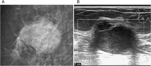
Figure 1 Image of a 65-year-old female patient in the PIMPC group. (A) The X-ray shows a high-density oval mass in the upper outer quadrant of the right breast with infiltrative margins and the burr sign. (B) The ultrasound shows a hypoechoic mass with uneven internal echoes and slightly reduced posterior echoes. The margin exhibits a crab claw-like pattern. The pathologic diagnosis was invasive micropapillary carcinoma of the right breast, 1.8 cm×1.5 cm×1.2 cm in size.
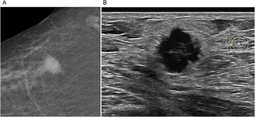
Figure 2 Image of a 72-year-old female patient. (A) The X-ray shows a high-density oval mass in the upper outer quadrant of the right breast with infiltrative margins and no burr sign. (B) The ultrasound shows a hypoechoic mass with uneven internal echoes and slightly reduced posterior echoes. The pathologic diagnosis was a non-specific invasive ductal carcinoma of the right breast (grade II), 2.8 cm×2.5 cm×2 cm in size.