Figures & data
Figure 1 Classification used and proposed by authors.
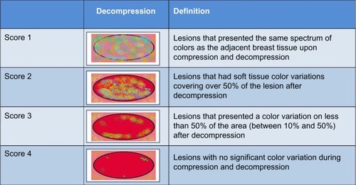
Table 1 Distribution of histopathologic results by sonoelastographic scores (%)
Figure 2 Age distribution for false-negative (group 1) and true-positive (group 2) results.
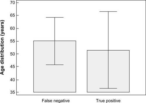
Table 2 Chi-square test to verify association between sonoelastographic scores and histologic groups
Figure 3 Size distribution for false-negative (group 1) and true-positive (group 2) results.
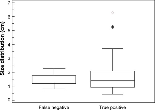
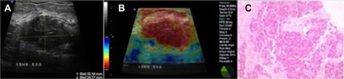
Figure 4 Atypical presentation of invasive ductal carcinoma in a 40-year-old patient, presenting as a palpable mass.
Abbreviation: BI-RADS, Breast Imaging and Reporting Data System.
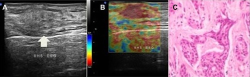
Figure 5 Heterogeneous mass in the left breast of a 45-year-old patient.
Abbreviation: BI-RADS, Breast Imaging and Reporting Data System.
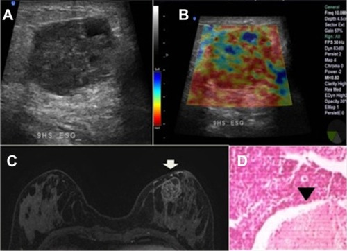
Figure 6 Clustered retroareolar microcalcifications in the right breast of a 52-year-old patient.
Abbreviation: BI-RADS, Breast Imaging and Reporting Data System; CCD, right craniocaudal view.
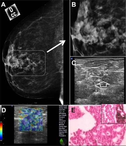
Figure 7 Mucinous carcinoma in a 58-year-old patient.
Abbreviation: BI-RADS, Breast Imaging and Reporting Data System; CCD, right craniocaudal view; CCE, left craniocaudal view.
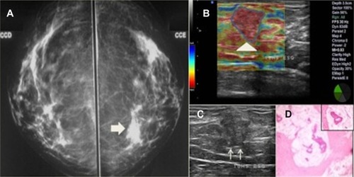
Figure 8 Invasive ductal carcinoma of intermediate grade in a 63-year-old patient.
Abbreviation: BI-RADS, Breast Imaging and Reporting Data System.