Figures & data
Figure 1 Histopathological sections of the original tumor of which the AMJ13 cell line was derived from.
Notes: (A) A duct containing finger-like projections covered by a layer of epithelial and underlying myoepithelial cells with a fibrous core (hematoxylin and eosin staining, magnification 40×). (B) Infiltrating ductal carcinoma composed of irregular solid groups of cells in a dense fibrous stroma (hematoxylin and eosin staining, magnification 40×).
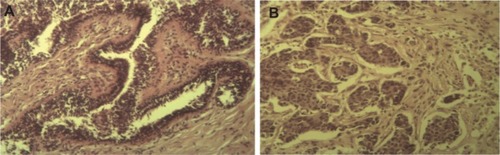
Figure 2 AMJ13 cell line morphology.
Notes: (A) Morphology of AMJ13 cells in culture showing (B) an epithelial-like cell shape with multiple nuclei, a high N/C ratio, and the presence of mitotic figures (hematoxylin and eosin staining, magnification 40×). (C) Scanning electron micrographs of human breast cancer-derived cell cultures. The cells are squamous with many short and thin processes and grow upon each other (magnification 1,000×). (D) Scanning electron photomicrograph showing AMJ13 cells with characteristic epithelial polygonal shape (magnification 3,000×) (E) Scanning electron photomicrograph showing microvilli on the cancer cells that ranged from 0.792 µm to 5.98 µm in length (magnification 10,000×). (F) Scanning electron photomicrograph showing the measurements for AMJ13 cancer cells, for which the average length was 73.8 µm, the average width was 33 µm, and the average nucleus diameter was 11.1 µm (magnification 1,000×).
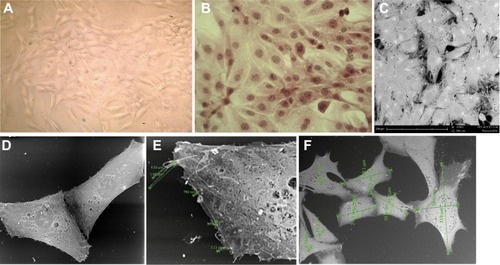
Figure 3 Growth curve for AMJ13 cell line, with a population doubling time of 22 hours.
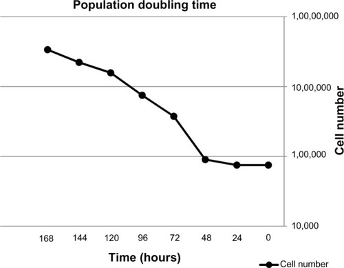
Figure 4 AMJ13 cells growing in an anchorage-independent fashion using soft agar colony assays.
Notes: (A) AMJ13 cells readily formed colonies (93.85 µm in diameter) within 6 days. (B) After 14 days, the colonies continue to expand, reaching 282.75 µm in mean diameter. This result demonstrates that AMJ13 cells undergo efficient anchorage-independent growth.
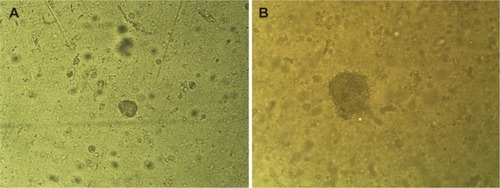
Figure 5 Analysis of Karyotype.
Notes: (A) Representative metaphases. (B) G band karyotype for the established breast cancer cell line that show most chromosomes as markers.
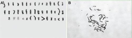
Figure 6 Chromosome numbers differ between cells.
Notes: (A) disrupted metaphase with 33 chromosome, (B) disrupted metaphase with chromosome number more than 47, and (C) disrupted metaphase with chromosome number less than 30.
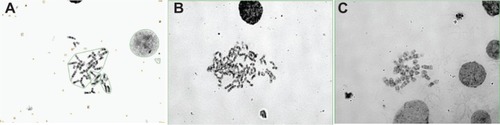
Figure 7 Histogram showing abnormal chromosome numbers in AMJ13 cells.
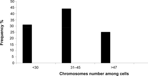
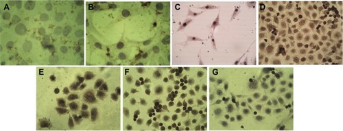
Figure 8 Immunocytochemistry analysis of AMJ13 cell line.
Notes: (A) Negative result for ER (magnification 40×). (B) Negative result for PR (magnification 40×). (C) Weak positive result for Her-neu2 gene expression (magnification 40×). (D) BRCA1-positive nuclear staining of breast cancer epithelial cells (magnification 40×). (E) BRCA2-positive nuclear and cytoplasmic staining of breast cancer cells (magnification 40×). (F) Vimentin marker-positive cells (magnification 40×). (G) Negative control.