
Figures & data
Figure 1 Planning of the pedicle.
Notes: Black arrow points to the part to be resected; red arrow points to the intact part of the pedicle; blue arrow points to the deepithelialized part of the pedicle; green arrow points to the tumor location marked after ultrasonographic guidance.
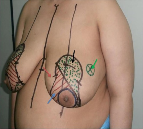
Figure 2 Tumor resection after deepithelialization.
Notes: (A) Deepithelialization of the distal half of the pedicle; black arrow points to the part to be resected; red arrow points to the intact part of the pedicle; blue arrow points to the deepithelialized part of the pedicle; (B and C) excision of the tumor-bearing area; (D) appearance after removal of the tumor-bearing area; (E) axillary dissection from the same wound; (F) specimen containing the tumor.
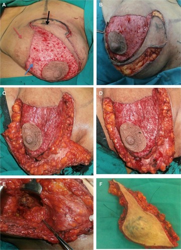
Figure 3 Therapeutic mammoplasty.
Notes: (A) One-month postoperative view after bilateral medial pedicle therapeutic mammoplasty; (B) 3 months after unilateral procedure.
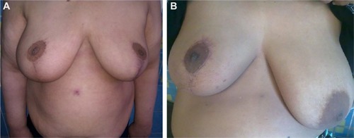
Table 1 Tumor characteristics
Table 2 Postoperative complications