Figures & data
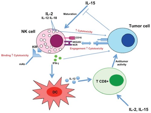
Figure 1 Electron micrographics of natural killer (A) and NK-92 (B) cells showing large lymphocyte-containing granules (arrows).
Abbreviation: N, nucleus.
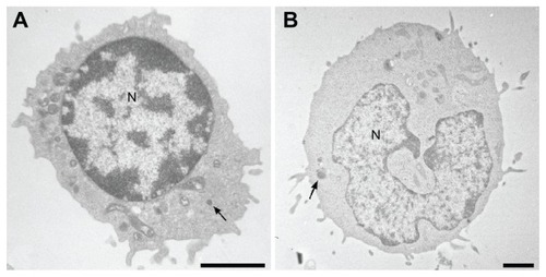
Figure 2 Recognition mechanisms of target cells by NK cells: “missing and induced self” theory. NK cell response is not initiated if neither ligands for NK-activating receptors nor MHC-I are expressed on target cells (A). If inhibitory receptors interact with MHC-I molecules without ligands for activating receptors no cytotoxicity is observed (B), whereas engagement of these receptors in absence of MHC-I molecule induced a strong NK cell response (C). In most cases, NK cell response depends on a balance between inhibitory and activating receptor signaling (D). Normal cells are protected against NK cell cytotoxicity because they usually express MHC-I molecules and no or low level of activating receptor ligands.
Abbreviations: MHC-I: major histocompatibility complex class I; NK, natural killer.
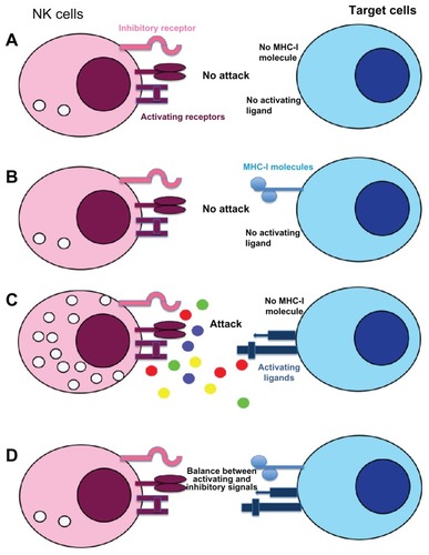
Figure 3 Human natural killer cell subsets based on CD56 and CD16 expressions: Around 90% of natural killer cells isolated from the blood display dim level of CD56 and high density of CD16 (CD56dimCD16bright).
Abbreviations: GM-CSF, granulocyte–macrophage colony-stimulating factor; IFN-γ, interferon γ; IL-10, interleukin 10; TNF-α, tumor necrosis factor α.
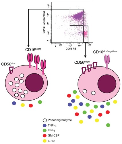
Table 1 Main receptors on NK cells involved in anti-tumor immune response
Figure 4 Overview of NK cell responses against tumor cell.