Figures & data
Figure 1 Design structure of the Promus Element stent.
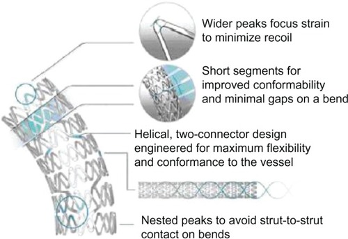
Figure 2 Key differences between the Promus Element and Synergy stent. In the thinner-strut Synergy stent, the drug and bioabsorbable poly-DL-lactide-co-glycolide (PLGA) polymer are applied to the abluminal stent surface only (A). The Synergy stent has different strut thickness, connector angle, and peak radius diameters, resulting in an enhanced stent platform (B). Panel (C) shows similar radiopacity between the stents despite reduced strut thickness in the Synergy stent.
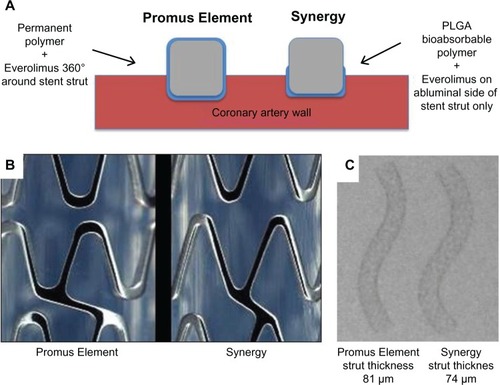
Figure 3 Kinetics of drug release and polymer absorption with the Synergy stent.
Abbreviation: PLGA, poly-DL-lactide-co-glycolide.
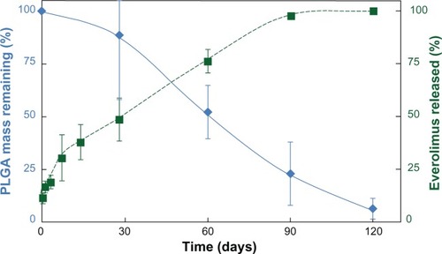
Table 1 PLATINUM and EVOLVE Clinical Trial Programs
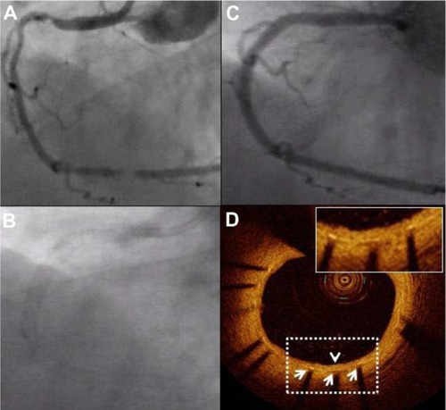
Figure 4 Coronary angiography and optical coherence tomography (OCT) 9 months after Promus Element implantation. A diffusely diseased right coronary artery (A) was treated with three slightly overlapping Promus Element stents. Panel (B) confirms excellent radiopacity of the Platinum Chromium Element stent platform. Follow-up angiography at 9 months shows prolonged vessel patency (C). A representative OCT image confirms favorable healing: all struts (white arrows) are covered with a thin homogeneous layer of neointimal tissue (arrowhead) with a well-preserved lumen area (D).