Figures & data
Figure 1 Schematic diagram of the process of erythropoiesis. The various stages of erythroid differentiation are shown including the key cytokines that are involved in the proliferation, survival and differentiation of the erythroid progenitors.
Abbreviations: BFU-E, burst forming unit-erythroid; CFU-E, colony forming unit-erythroid; CFU-GEMM, colony forming unit-granulocyte, erythroid, macrophage, megakaryocyte; EPO, erythropoietin; G-CSF, granulocyte colony stimulating factor; GM-CSF, granulocyte monocyte colony stimulating factor; IL-3, interleukin 3; IL-9, interleukin 9, IGF-1, insulin-like growth factor 1.
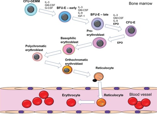
Figure 2 In vitro proliferation activity and EPOR binding activities of rHuEPO, darbepoetin alfa, AMG 114, and AMG 205 glycosylation analogs.
Notes: (A) HCitation3-thymidine incorporation assays in UT-7/EPO cells demonstrates the inverse correlation between molecules with increasing numbers of N linked carbohydrate (rHuEPO = 3; darbepoetin alfa = 5; AMG 114 and AMG 205 = 7) and decreasing in vitro proliferation activity. Proliferation is measured as counts CPM of HCitation3-thymidine incorporation into newly synthesized DNA and EC50 values of a representative experiment are shown. (B) Competitive binding assays were performed in UT-7/EPO cells whereby I125-rHuEPO was bound to cells and competed with increasing concentration of ESA for 2–3 hours. Cells were washed though phthalate oil, CMP was measured and IC50 calculated. A representative experiment is shown. Data kindly provided by Steve Elliott, Norma Rogers, and Tony Lorenzini, Amgen, Inc.
Abbreviations: CPM, counts per minute; EC50, 50% effective concentration; IC50, 50% inhibitory concentration; EPO, erythropoietin; EPOR, EPO receptor; rHuEPO, recombinant human EPO.
Abbreviations: CPM, counts per minute; EC50, 50% effective concentration; IC50, 50% inhibitory concentration; EPO, erythropoietin; EPOR, EPO receptor; rHuEPO, recombinant human EPO.
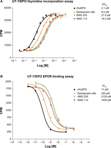
Table 1 Summary table of the in vitro proliferation EC50 values and the IC50 receptor binding activity in rHuEPO competition assays using UT-7/EPO cells
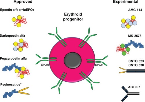
Figure 3 Schematic representation of approved and experimental ESA molecules.
Notes: Shown are schematic diagrams of the molecular structures: red molecule, EPO-based peptide; yellow circles, carbohydrate attached to naturally occurring N-linked glycosylation sites; light blue circles, carbohydrate attached to engineered N-linked glycosylation sites; green circles; carbohydrate generated in yeast at naturally occurring N-linked glycosylation sites; small blue circles, PEG; orange ovals, EPO mimetic peptides; dark blue rectangles, antibodies or Fc portion of antibody. An erythroid progenitor with surface EPOR is also shown. *As of February 2013, peginesatide is no longer on the market.
Abbreviations: EPO, erythropoietin; EPOR, EPO receptor; ESA, erythropoiesis stimulating agent; PEG, methoxy-polyethylene glycol; rHuEPO, recombinant human EPO.
Abbreviations: EPO, erythropoietin; EPOR, EPO receptor; ESA, erythropoiesis stimulating agent; PEG, methoxy-polyethylene glycol; rHuEPO, recombinant human EPO.