Figures & data
Figure 1 Schematic of the influenza HA epitope region and peptide sequences from the current study.
Notes: The position of the upper and lower peptides in the primary sequence of HA1 and their amino acid sequences are indicated. F’, E’, and R (A). Overlapping 15-mer peptides derived from the upper (U-A, U-B, U-C, and U-D) and lower (L-A and L-B) regions that were used to examine antibody specificity (B). Image of the HA molecule showing the position of the upper (red) and lower (green) epitope peptide regions (C). The images were created by PyMol (DeLano Science) using the HA structure obtained from the Protein Data Bank.Citation14 The monomer HA was created using accession number 2VIU, described by Fleury et al, and the trimer HA was created using accession number 1HGI, described by Sauter et al.Citation14 The upper and lower peptides form an antiparallel β-sheet structure. Large portions of the N-terminus of the upper peptide and the C-terminus of the lower peptide are exposed on the surface underneath the receptor binding site, and small portions of the C-terminus of the upper peptide and the N-terminus of the lower peptide are exposed on the surface near the receptor binding site.
Abbreviations: F’, fusion peptide subdomain; E’, vestigial enzyme subdomain; R, receptor binding subdomain; HA, influenza hemagglutinin; N, N-terminal portion of the peptide; C, C-terminal portion of the peptide.
Abbreviations: F’, fusion peptide subdomain; E’, vestigial enzyme subdomain; R, receptor binding subdomain; HA, influenza hemagglutinin; N, N-terminal portion of the peptide; C, C-terminal portion of the peptide.
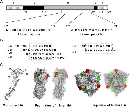
Figure 2 Binding activity of different rabbit antisera to epitope peptides, as determined by ELISA.
Notes: N-terminally biotinylated upper peptide (A and B); N-terminally biotinylated lower peptide (C and D); C-terminally biotinylated upper peptide (E and F); and C-terminally biotinylated lower peptide (G and H) were immobilized via biotin–streptavidin binding onto an ELISA plate that was precoated with streptavidin. The antisera were serially diluted from 1:1,000 to 1:125,000. Rabbits immunized are designated as aU-1, aU-2, aL-1, aL-2, aU+L-1, and aU+L-2. Individual rabbits are indicated by “-1” and “-2.” Experiments were carried out in duplicate, and the results are presented as means.
Abbreviations: aU, rabbit immunized with upper peptide alone; pre, serum harvested from each rabbit prior to immunization; n, number; aU+L, rabbit immunized with both the upper and lower peptides; aL, rabbit immunized with lower peptide alone; ELISA, enzyme-linked immunosorbent assay.
Abbreviations: aU, rabbit immunized with upper peptide alone; pre, serum harvested from each rabbit prior to immunization; n, number; aU+L, rabbit immunized with both the upper and lower peptides; aL, rabbit immunized with lower peptide alone; ELISA, enzyme-linked immunosorbent assay.
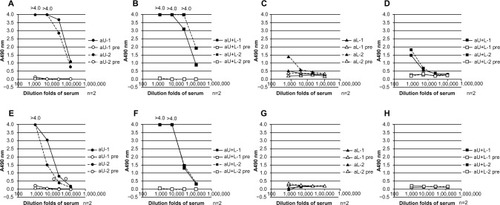
Figure 3 Binding ability of different rabbit antisera to HA, determined by ELISA and immunoblotting.
Notes: The HA vaccine preparation was used as coating antigen for an ELISA. The rabbit antisera were diluted serially from 1:1,000 to 1:25,000. Experiments were carried out in duplicate and results are presented as means (A, B, and C). For immunoblotting, different influenza HA vaccine antigens were subjected to SDS-PAGE, and immunoglobulin G purified with Protein-G® Sepharose (GE Healthcare UK Ltd, Little Chalfont, UK) from the antisera of rabbits aU-1, aU+L-1, and aU+L-2 was used as the probe at a concentration of 1 μg/mL or 5 μg/mL. Individual rabbits are indicated by “-1” and “-2.” The designations “ ×1,” “ ×2,” and “ ×4” indicate that samples were loaded onto the gel at a one-, two-, or four-fold dilution.
Abbreviations: n, number; aU, rabbit immunized with upper peptide alone; pre, serum harvested from each rabbit prior to immunization; aL, rabbit immunized with lower peptide alone; aU+L, rabbit immunized with both the upper and lower peptides; Bri, influenza hemagglutinin vaccine antigen derived from A/Brisbane/59/2007 (H1N1); Urg, influenza hemagglutinin vaccine antigen derived from A/Uruguay/716/2007 (H3N2); Flo, influenza hemagglutinin vaccine antigen derived from B/Florida/4/2006; HA, influenza hemagglutinin; ELISA, enzyme-linked immunosorbent assay; SDS-Page, sodium dodecyl sulfate polyacrylamide gel electrophoresis.
Abbreviations: n, number; aU, rabbit immunized with upper peptide alone; pre, serum harvested from each rabbit prior to immunization; aL, rabbit immunized with lower peptide alone; aU+L, rabbit immunized with both the upper and lower peptides; Bri, influenza hemagglutinin vaccine antigen derived from A/Brisbane/59/2007 (H1N1); Urg, influenza hemagglutinin vaccine antigen derived from A/Uruguay/716/2007 (H3N2); Flo, influenza hemagglutinin vaccine antigen derived from B/Florida/4/2006; HA, influenza hemagglutinin; ELISA, enzyme-linked immunosorbent assay; SDS-Page, sodium dodecyl sulfate polyacrylamide gel electrophoresis.
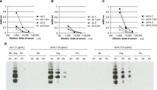
Figure 4 Binding abilities of antibodies affinity-purified with peptides to the HA vaccine, as determined by ELISA.
Notes: Antipeptide antibodies were purified by affinity binding with upper peptide immobilized on Sepharose via its N-terminus (A) or C-terminus (B), or with lower peptide immobilized on Sepharose via its N-terminus (C). Antibodies were diluted from 10 μg/mL to 0.1 μg/mL. Individual rabbits are indicated by “-1” and “-2.” Experiments were carried out in duplicate and results are presented as means. Normal IgG was purified from commercial normal rabbit serum (CEDARLANE, Burlington, ON, Canada) with Protein G Sepharose® (GE Healthcare UK Ltd, Little Chalfont, UK).
Abbreviations: aU, rabbit immunized with upper peptide alone; aU+L, rabbit immunized with both the upper and lower peptides; igg, immunoglobulin G; n, number; aL, rabbit immunized with lower peptide alone; HA, influenza hemagglutinin; ELISA, enzyme-linked immunosorbent assay.
Abbreviations: aU, rabbit immunized with upper peptide alone; aU+L, rabbit immunized with both the upper and lower peptides; igg, immunoglobulin G; n, number; aL, rabbit immunized with lower peptide alone; HA, influenza hemagglutinin; ELISA, enzyme-linked immunosorbent assay.
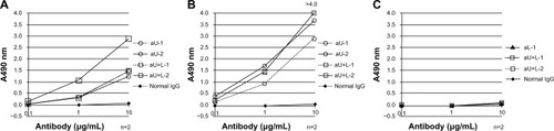
Figure 5 Binding affinities to recombinant HA, as determined by ELISA.
Notes: The ability of antisera (A) and affinity-purified antibodies (B) to bind recombinant HA was determined by ELISA. Antisera and affinity-purified antibodies were diluted to 1:1,000 and to 1 μg/mL, respectively. Experiments were carried out in duplicate and the results are presented as means (A). Experiments were carried out in triplicate and results are presented as means ± SD (B). Individual rabbits are indicated by “-1” and “-2.” Anti-upper-N or anti-upper-C antibody was produced by affinity-purified antisera with the upper peptide immobilized at the C-terminus or at the N-terminus, as described in . Sequence comparison of the upper and lower peptide between NY, which was used as the source of recombinant HA, and Urg, which was used as the vaccine preparation (C).
Abbreviations: aU, rabbit immunized with upper peptide alone; aL, rabbit immunized with lower peptide alone; aU+L, rabbit immunized with both the upper and lower peptides; n, number; Upper-C, C-terminal region of the upper peptide; Upper-N, N-terminal region of the upper peptide; SD, standard deviation; Urg, A/Uruguay/716/2007; NY, A/New York/55/2004; HA, influenza hemagglutinin; ELISA, enzyme-linked immunosorbent assay.
Abbreviations: aU, rabbit immunized with upper peptide alone; aL, rabbit immunized with lower peptide alone; aU+L, rabbit immunized with both the upper and lower peptides; n, number; Upper-C, C-terminal region of the upper peptide; Upper-N, N-terminal region of the upper peptide; SD, standard deviation; Urg, A/Uruguay/716/2007; NY, A/New York/55/2004; HA, influenza hemagglutinin; ELISA, enzyme-linked immunosorbent assay.
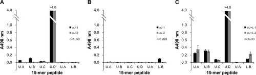
Figure 6 Binding activity of different rabbit antisera to 15-mer peptides within the upper and lower peptide regions of HA, as determined by ELISA.
Notes: Antisera from immunized rabbits were diluted 1:1,000 for use in the assay. The binding activity of antisera from aU rabbits (A), aL rabbits (B), and aU+L rabbits (C) was measured by ELISA. “U-A,” “U-B,” “U-C,” and “U-D” are 15-mer peptides located within the upper peptide region of HA, and “l-a” and “L-B” are 15-mer peptides within the lower peptide region of HA, as described in . Experiments were carried out in triplicate and results are presented as means ± SD.
Abbreviations: n, number; SD, standard deviation; HA, influenza hemagglutinin; ELISA, enzyme-linked immunosorbent assay; aU, rabbit immunized with upper peptide alone; aL, rabbit immunized with lower peptide alone; aU+L, rabbit immunized with both the upper and lower peptides.
Abbreviations: n, number; SD, standard deviation; HA, influenza hemagglutinin; ELISA, enzyme-linked immunosorbent assay; aU, rabbit immunized with upper peptide alone; aL, rabbit immunized with lower peptide alone; aU+L, rabbit immunized with both the upper and lower peptides.