Figures & data
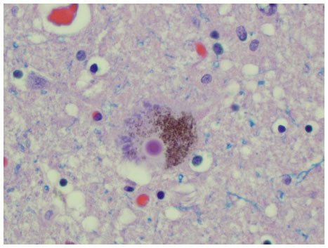
Figure 1 The central panel lists cellular pathways disrupted in the pathogenesis of PD that have been targeted for development of PD biomarkers. These abnormalities are thought to arise from a combination of genetic and environmental factors, as depicted on the left side of the panel.
Abbreviation: PD, Parkinson’s disease.
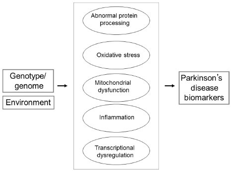
Table 1 Promising Parkinson’s disease-associated biomarkers in blood and CSF
Figure 2 Photomicrograph of a hematoxylin-eosin-stained section taken from the substantia nigra pars compacta at autopsy of an individual with PD. A dopaminergic neuron in the center of the field contains a densely stained cytoplasmic Lewy body.
Note: This article was published in Handbook of the Neuroscience of Aging. 2009, Schwarz C, Henchcliffe C. Parkinsonian syndromes. In: Hof P, Mobbs C, Editors. 441–447. Copyright ©2009 Academic Press.Citation2