
Figures & data
Figure 1 Painting “Greenland Mother” by W Langdon Kihn (1898–1957), showing a Greenlandic Inuit woman holding her baby. Note that her hair is worn in a tight coiffure arranged at the top of her head. Traumatic marginal alopecia can be appreciated, as was described by Trebitsch in 1907 as “alopecia Groenlandica.” From: National Geographic, October 1949, page 486. Public access.
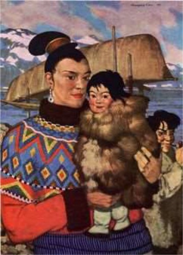
Table 1 Summary of traumatic hairstyles and their associated patient populations
Figure 2 Biopsy-proven traction alopecia involving the frontal and parietal scalp.
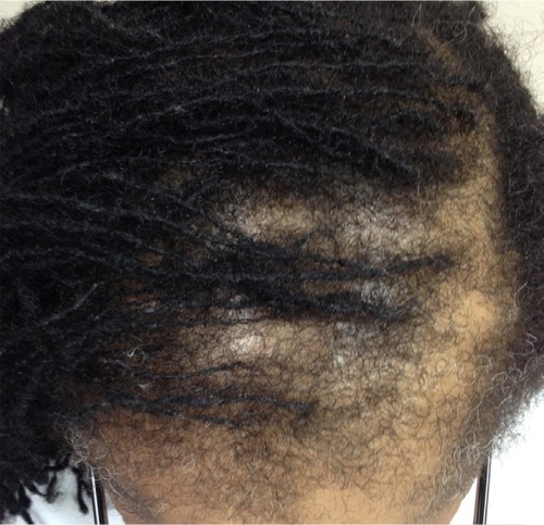
Figure 3 Marginal traction alopecia in patient with braids.
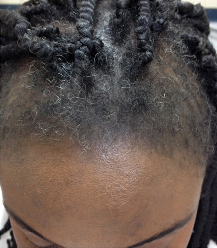
Figure 4 Trichoscopy of alopecic patch in traction alopecia shows only individual hairs with preserved marking of follicular openings outlined in brown that correspond to the pigmented basal cell layer of the infundibula.
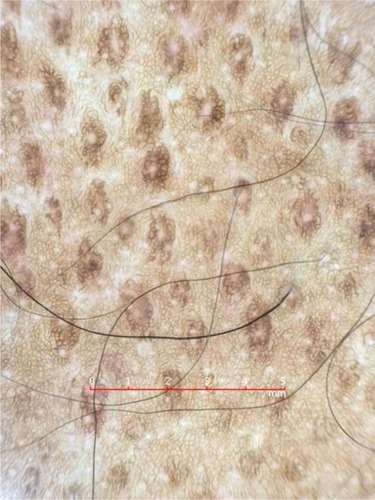
Figure 5 Trichoscopy of acute traction alopecia demonstrates black dots and broken hairs at different length. In this example, the patient is presented with marginal hair loss after removal of a glued wig.
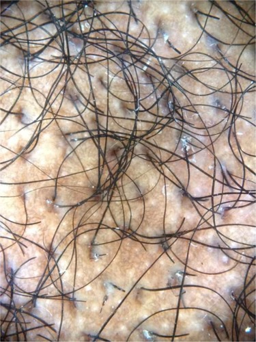
Figure 6 Trichoscopy of traction alopecia demonstrates cylindrical hair casts along several hair shafts.
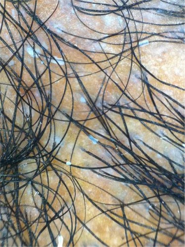
Figure 7 Histology of traction alopecia. On horizontal sections at the level of the low follicular level, there is altered follicular architecture due to areas of follicular drop out. There are only four follicles (hematoxylin and eosin, ×2).
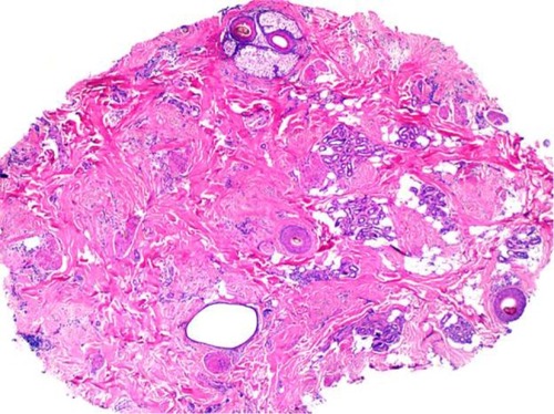
Figure 8 Histology of traction alopecia. On horizontal sections at the level of the isthmus, there is an overall preserved follicular architecture but significantly reduced follicular density (total of nine follicles). The terminal: vellus ratio is 2:1. The sebaceous glands are mostly preserved (hematoxylin and eosin, ×2).
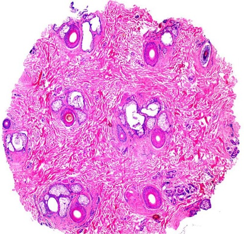
Table 2 Summary of the distinguishing clinical, trichosopic, and pathologic findings in patients with patchy alopecias