Figures & data
Table 1 Case studies claiming delusional etiology of Morgellons disease
Figure 1 Embedded cutaneous blue and white filaments.
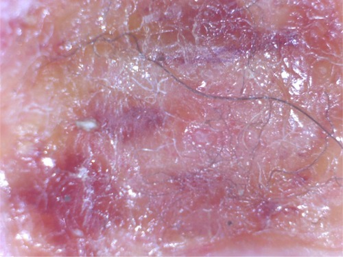
Figure 2 Longitudinal sections of filaments originating in the basal layer of the epidermis adjacent to the dermis; magnification 400×.
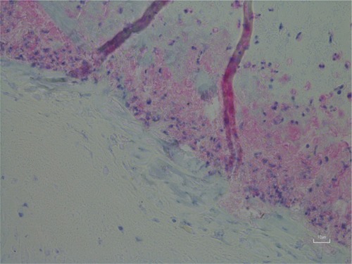
Figure 3 Filaments remaining embedded in deeper layers of skin after removal of a callus; magnification 100×.
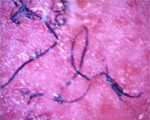
Figure 4 (A) A filamentous follicular cast. White filaments originating on the outer follicular sheath are growing in a coiled manner. Magnification 50×. (B) Pili multigemini, a common finding in Morgellons disease patients, with multiple hairs forming from a single bulb. Magnification 50×.
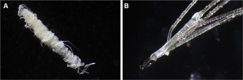
Figure 5 (A) Thickened keratinized follicular casts in a Morgellons disease specimen that grew inward into the dermis. Note the clear inward-growing hair. Magnification 100×. (B) Specimen from a bovine digital dermatitis lesion with similarities to human Morgellons specimens. Note thickened keratin projections and the threadlike blue filament (lower part of specimen). Magnification 50×.
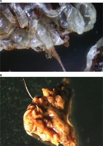
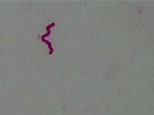
Figure 6 Single spirochete from a Morgellons disease skin specimen immunostained for detection of Borrelia. Magnification 1,000×.