
Figures & data
Figure 1 Flow chart of inclusion and exclusion criteria and final selection of rhinophyma.
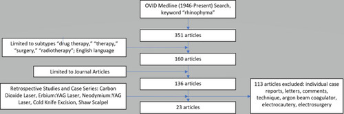
Table 1 Classification and Grading of Rhinophyma
Table 2 Variants of Rhinophyma
Table 3 Post-Operative Outcome Scales
Table 4 Surgical Outcomes by Study
Table 7 Summary of Outcomes by Treatment Type
Table 8 Summary of Complications by Treatment Type
Table 5 Surgical Outcomes by Study
Table 6 Summary of Outcomes by Treatment Type
Figure 2 Subunit approach for refractory rhinophyma. (A) Intra-operative markings with six subunit based flaps (dorsum, two sidewalls, two alae, tip). (B) Subunit flaps are raised to provide exposure for removal of the phymatous tissue and facilitate correction of nasal support with cartilage grafts. (C) Extensive debulking of phymatous tissue to perichondrium. (D) Excess skin that has been “expanded” by sebaceous growth is trimmed.
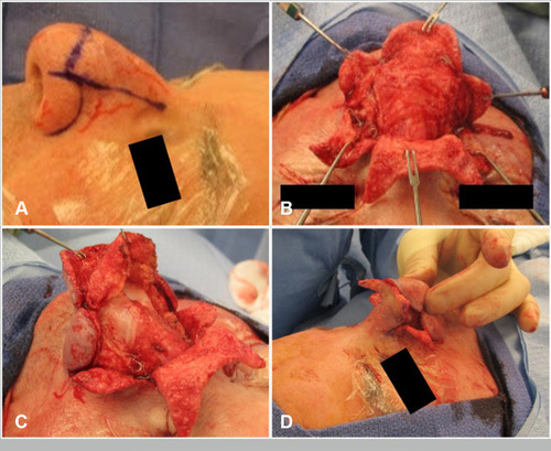
Figure 3 (A) Pre-operative photo of patient with rhinophyma refractory to laser and tangential excision. (B) Intra-operative result after undergoing subunit method. Note incisions are planned at the subunit junctions. (C) Post-operative image with improvement in rhinophyma and nasal definition.
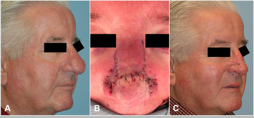
Table 9 Summary of Treatment Advantages and Disadvantages