
Figures & data
Figure 1 Microscope images of: (A) translucent, non-pigmented, septate hyaline hyphae of Trichophyton rubrum (blue arrow) in a nail sample (10% KOH stain, ×400). (B) Endothrix hair invasion, with translucent and non-pigmented spores of Trichophyton violaceum (10% KOH stain, ×100). (C) Bluish, narrow septate hyphae of Trichophyton rubrum against a pink background in a nail sample (Chicago sky blue stain, ×400). (D) Endothrix hair invasion with blue spores of Trichophyton violaceum (Chicago sky blue stain, ×100).
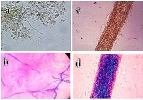
Figure 2 (A) Endothrix hair invasion with green, fluorescent hyphae and Trichophyton violaceum spores (Calcoflour white stain, fluorescent microscope, ×400). (B) Fluorescent septate hyphae of Trichophyton rubrum against a darker background in a nail sample (Calcoflour white stain, fluorescent microscope, ×100). (C) Anterior surface of a Trichophyton rubrum culture. (D) Reverse surface of a Trichophyton rubrum culture. (E) Microscope image of blue, thin walled Trichophyton rubrum macroconidia showing the clavate shape and multiseptate, smooth walled structure (lacto phenol cotton blue stain, ×100).
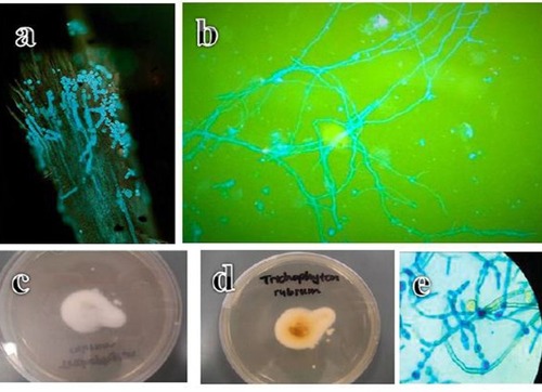
Table 1 Results Of Chicago Sky Blue And Calcoflour White Stains In Comparison With KOH Wet Mount And Culture On Sabouraud’s Dextrose Agar In Onychomycosis
Table 2 Results Of Chicago Sky Blue And Calcoflour White Stains In Comparison With KOH Wet Mount And Culture On Sabouraud’s Dextrose Agar In Tinea Capitis