
Figures & data
Table 1 Demographic and Clinical Data of Studied Subjects
Figure 1 (A) Patchy mild cytoplasmic expression of gal-9 (red boxes) in control epidermis (Immunoperoxidase, 400 ×); (B) Diffuse moderately nucleo-cytoplasmic expression (red arrows) of gal-9 is in epidermal keratinocytes in AD skin section (Immunoperoxidase, 40 ×); (C) Mild nuclear expression (red arrows) of gal-9 in the epidermal keratinocytes of AD skin section (Immunoperoxidase, 400 ×).
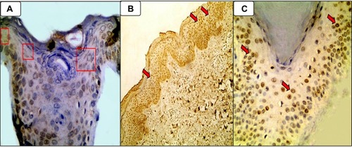
Table 2 Comparison Between Studied Groups Regarding Galectin-9 Immunoreactivity
Figure 2 (A) Mild focal cytoplasmic expression of E selectin (red arrows) in epidermal keratinocytes of control section (Immunoperoxidase, 400 ×); (B) Moderate focal cytoplasmic expression of E selectin (red arrows) in AD epidermal keratinocytes (Immunoperoxidase, 200 ×); (C) Diffuse moderate predominantly nuclear expression of E selectin (red circles) in the epidermal keratinocytes of AD skin section (Immunoperoxidase, 400 ×).
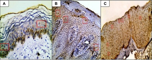
Table 3 Comparison Between Studied Groups Regarding E Selectin Immunoreactivity
Figure 3 Showing co-localization of the both E-selectin (A) and galectin-9; (B) immunohistochemical nucleo-cytoplasmic expression (red circles in both (A and B) in the epidermis of a case of atopic dermatitis (immunoperoxidase 200 × HPF [A], 40 × HPF [B]).
![Figure 3 Showing co-localization of the both E-selectin (A) and galectin-9; (B) immunohistochemical nucleo-cytoplasmic expression (red circles in both (A and B) in the epidermis of a case of atopic dermatitis (immunoperoxidase 200 × HPF [A], 40 × HPF [B]).](/cms/asset/23d7ab47-264b-4954-8743-8342b46ea4fe/dcci_a_12161596_f0003_c.jpg)
Table 4 Relationship Between Galectin-9 H Score and Studied Parameters