
Figures & data
Figure 1 (A and B) Small porcelain-white papules and macules with peripheral telangiectasia. (C) Multiple atrophic porcelain-white macules with peripheral telangiectasia on reticulated erythematous to brownish background.
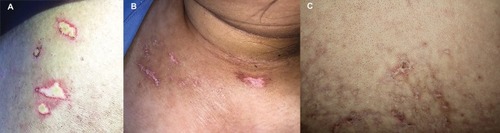
Figure 2 (A) Histopathology from case 1 showed hyperkeratotic epidermal atrophy, vacuolar alteration of basal keratinocytes, marked papillary edema with extravasated red blood cells, and telangiectasia (H&E, 100x). (B) Histopathology from case 3 showed wedge-shaped infarct in papillary dermis with sparse lymphoplasmacytic infiltrate and vacuolar alteration of basal cell layer. Homogenized eosinophilic altered collagen bundles within the entire dermis (H&E, 100x).
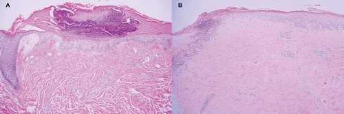
Figure 3 Direct immunofluorescence shows epidermal nuclear staining of immunoglobulin G (400x).
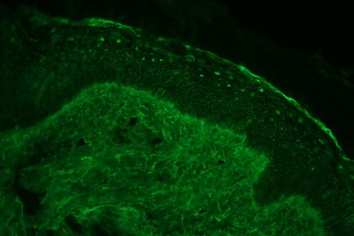
Table 1 Demographic Data, Clinical, Laboratory, Histopathologic, Immunopathologic Characteristics, Treatment, And Outcome Of Degos-Like Lesions In Patients With Connective Tissue Diseases