Figures & data
Figure 1 Photograph of the lesion on the left nipple. Erosion and crust on the left nipple.
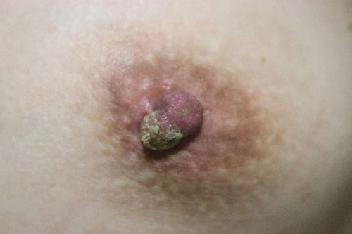
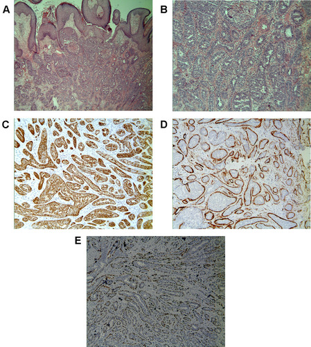
Figure 2 (A and B) Histopathology reveals ductal differentiation. (Hematoxylin-eosin stain, Original magnification×40 (A); Original magnification×200 (B)). The luminal epithelial cells were positive for (C) CK5/6 stain. (Immunohistochemistry, original magnification×100). The out-layer myoepithelial cells were positive for (D) SMA and (E) p63 stains. (Immunohistochemistry, original magnification×100).