
Figures & data
Figure 1 Frequency distribution of primary cutaneous lymphomas.

Figure 2 Primary cutaneous marginal zone lymphoma: clinical findings. (A) Male, 18 years old. Small erythematous papule on the volar surface of the left thigh. (B) Dermoscopically, the lesion appeared as a salmon‐coloured area with prominent blood vessels with serpentine (linear‐irregular) morphology.
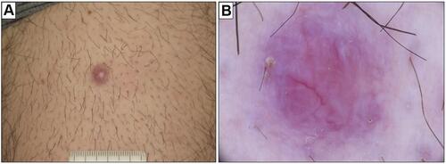
Figure 3 Primary cutaneous marginal zone lymphoma: histological findings. A diffuse lymphoid population in the reticular dermis, extending along a hair ((A) H&E, original magnification 20x). In the deeper dermis, some sweat glands are entrapped in the lymphoid infiltrate, in absence of lympho-epithelial lesions ((B) H&E, original magnification 40x). A granulomatous reaction is possible at the periphery of the infiltrate ((C) H&E, original magnification 40x). The lymphoid population is heterogeneous, including mature lymphocytes, lympho-plasmacytoid cells and plasma cells ((D) H&E, original magnification 200x).
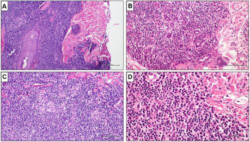
Table 1 Main Contribution of Immunohistochemical Markers to Diagnosis of PCMZL
Figure 4 Primary cutaneous marginal zone lymphoma: immunohistochemical findings. Lymphoid population showing positivity for CD20 and Bcl2. CD21 immunostaining highlights a partially destroyed network of follicular dendritic cells. An abundant reactive population of CD138-positive plasma cells is present at the periphery of the infiltrate. In-situ hybridization (ISH) demonstrates a kappa:lambda ratio of about 3:1.
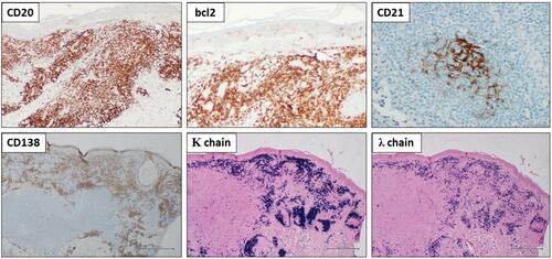
Table 2 ISCL/EORTC Staging System for Primary Cutaneous Lymphomas