Figures & data
Figure 1 Lentigo maligna melanoma on the scalp of a 78-year old man with androgenic alopecia (A–C) and solar lentigo on the scalp of a 81-year old man. (D–F) (A) Clinical appearance of the LMM. A large macule, variegated in color from light to dark brown to black with ill defined borders. In the background, signs of extensive sun-damage. (B) Dermoscopy showing a brown pseudo network with grayish circles around follicular openings and white streaks. (C) Dermoscopy of the hyper pigmented part, with obliteration of follicular openings, blue white veil and irregular dots. (D) Clinical appearance of a solar lentigo: a brown macule with a hyperkeratotic component. (E and F) Dermoscopy showing a light brown pseudo network with evident follicular openings and cerebriform pattern.
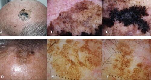
Figure 2 In situ melanoma of the vertex in a 68 year-old woman. (A) The lesion was arising in hair bearing scalp, the patient was not aware of the macule which was noticed by the hair dresser. (B) Close up clinical image, a flat brown macule 1 cm in diameter. (C and D) In dermoscopy atypical network and regression are detected. No non-prevalent benign pattern.
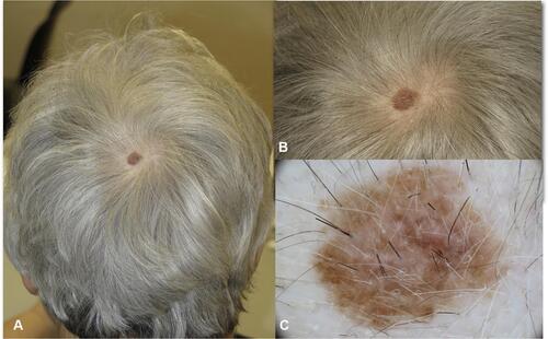
Figure 3 Superficial spreading melanoma on the scalp of a 70 year old man. (A) The lesion is arising in an area of thinning hair due to androgenic alopecia. (B) In dermoscopy, atypical network, irregular globules and blue-white veil are visible.
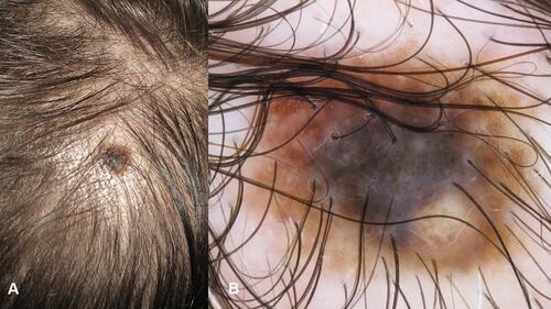
Figure 4 Nevus associated melanoma arising on the scalp in a 60-year old man with androgenic alopecia. (A) Clinically, a asymmetrically pigmented macule is visible surrounding a skin colored papule. (B) In dermoscopy, two components are visible, one flat with atypical pseudo network and obliteration of hair follicles, grey circles, black and brown dots. The pinkish area represents remnants of a dermal nevus.
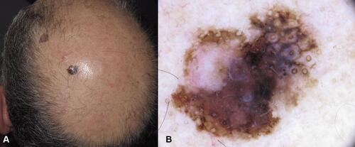
Figure 5 Nevus associated melanoma on the scalp of a 45-year old man. (A) An asymmetric lesion with brown and blue component. (B) In dermoscopy, on one side the congenital nevus with globular pattern is seen, on the other side the melanoma component with blue-white veil, shiny-white streaks.
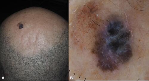
Figure 6 Scalp nevi in children. (A and B) brown pigmented macule on hair bearing scalp. (C) Regular globular pattern. (D) Targetoid pattern in a compound nevus.
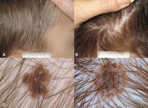
Figure 7 Scalp nevi in adults. (A–C) Clinical appearance of partially pigmented (A) and amelanotic (B and C) dome shaped papules. (D) In Dermoscopy, globular pattern in a compound nevus. (E and F) Dermoscopy of amelanotic lesions: blurred vascular pattern in two dermal nevi.
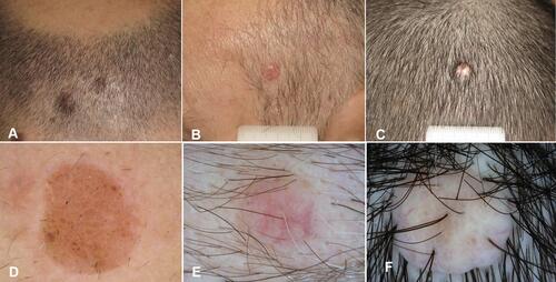
Figure 8 Blue nevus. (A) A blue papule on the frontal area of a 50-year old woman. (B) In dermoscopy structureless blue pigmentation is detected.
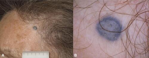
Figure 9 Invasive melanoma on the parietal region in a 75-year-old man with androgenic alopecia, superficial spreading melanoma 3.3 mm Breslow thickness (A) The lesion appears partially nodular with not-defined borders. (B, C) In dermoscopy a unspecific pattern is detected with brownish pigmentation, white lines and milky red areas.
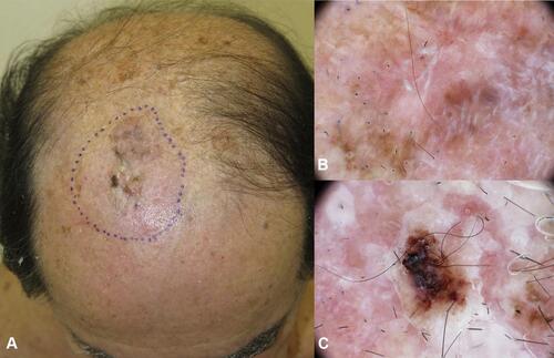
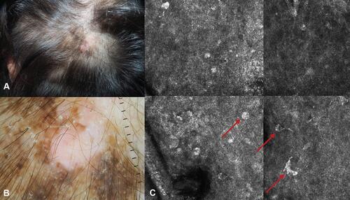
Figure 10 Pigmented basal cell carcinoma on the vertex in a 72-year old woman. (A) Clinically a heavily pigmented lesion with ill defined borders and superficial crusts. (B) Dermoscopy, highlighting the presence of black pigmentation and grey blue ovoid nests. (C) RCM. Confocal mosaic (2x1mm) at the level of the dermal epidermal junction showing multiple basaloid islands, visible as basaloid nests with palisading and clefting and the surrounding vascular structures.
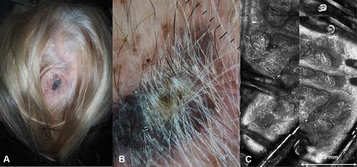
Figure 11 (A) Amelanotic nodular melanoma on the scalp of a 75-year old man, 4.5 mm Breslow thickness. (B) Close up of the non pigmented nodule. (C) In dermoscopy atypical vascular pattern is detected.
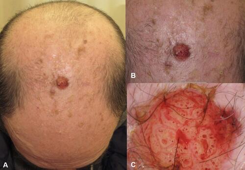
Figure 12 Desmoplastic melanoma on the scalp of a 68-year old woman. (A) Clinical appearance of the dome-shaped melanotic papule, with surrounding brownish macule. (B) The dome shaped papule corresponds to the invasive desmoplastic component, the brown-greyish macule with pseudo network corresponds to the lentigo maligna component. (C) RCM image highlighting the presence of atypical melanocytes (red arrows).