Figures & data
Figure 1 Ulcerated, punctate erosions with ragged edges on the patient’s back typical of Morgellons disease lesions.
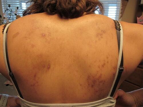
Table 1 Results of Laboratory Testing
Figure 2 (A–C) Expanding annular rashes on the patient’s hands, back and leg, with a raised, advancing, erythematous border consistent with secondary erythema migrans (EM) rashes.
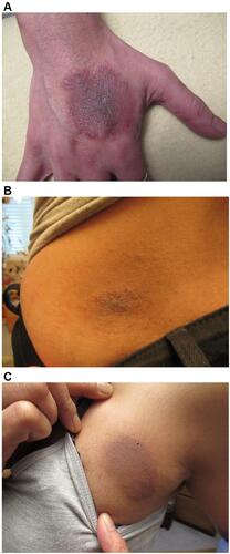
Figure 3 Secondary EM rash revealing embedded fibers (arrow) consistent with Morgellons disease. 100X original magnification.
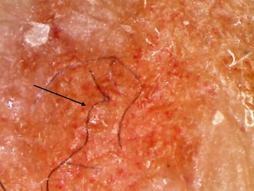
Figure 4 Gross morphology of a biopsy taken from the advancing edge of a lesion on the hand, revealing embedded and protruding blue and white fibers. 100X original magnification.
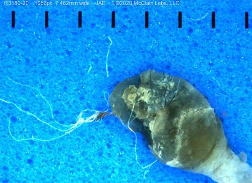
Figure 5 Sections stained with H&E exhibiting parakeratotic hyperkeratosis, spongiosis, and hemorrhage. 200X original magnification.
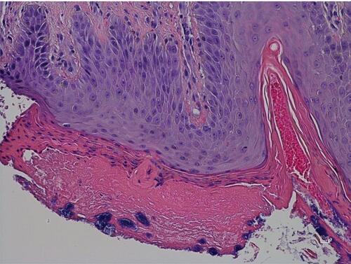
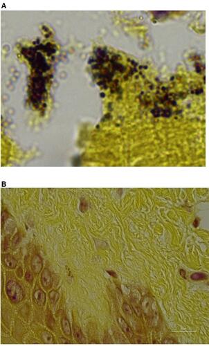
Figure 6 (A) Dieterle silver stain of spirochetes (arrow) among basal keratinocytes. 1000X original magnification. (B) Anti-Bb immunostained section showing positive-stained Borrelia organisms consistent with cystic morphology mixed with negatively stained bacteria within bacterial aggregates. 1000X original magnification.

Figure 7 (A) Anti-Bb immunostained section showing positively stained intracellular and extracellular organisms (arrow) associated with keratinocytes of the stratum basale and stratum spinosum. 1000X original magnification. (B) Anti-Bb immunostained section showing intracellular staining of organisms within lymphocytes (arrow) in a dermal infiltrate. 1000X original magnification. (C) Anti-Bb immunostained section showing positively stained organisms (arrow) in dermal glandular tissue. 1000X original magnification. (D) Anti-Bb immunostained control section of fungal-infected skin. 1000X original magnification.
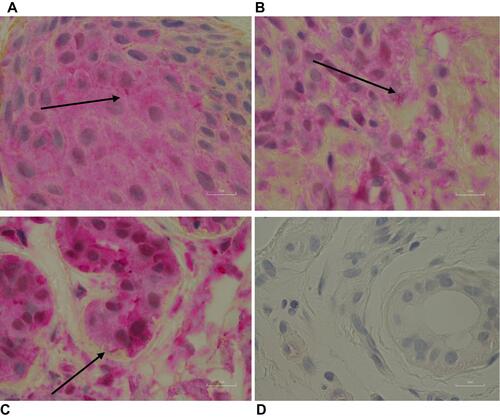
Figure 8 (A) Gram stain of biopsy section showing Gram-positive cocci in superficial layer. 1000X original magnification. (B) Gram stain of biopsy section showing negative staining in deep layers. 1000X original magnification.