
Figures & data
Table 1 Tear Trough Deformity Classification Systems.
Figure 1 Hirmand classification of tear trough deformity. Data from Hirmand.Citation5
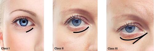
Figure 2 Overview of the hyaluronic acid (HA) injectable filler on the muscle activity. Data from de Maio.Citation11 Image courtesy from and reprinted with permission from Allergan plc, Dublin, Ireland.
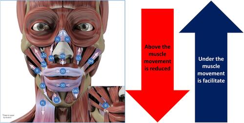
Table 2 Belhaouari et alCitation9 Tear Trough Classification System According to the Volume and the Ptosis with the Treatment Strategy with Hyaluronic Acid Filler
Table 3 Mechanic Myomodulation Effect with Hyaluronic Acid (HA) Filler on the Different Treatment Points.
Figure 3 Overview of the different codes involved in the tear trough deformity direct approach treatment. Image courtesy from and reprinted with permission from Allergan plc, Dublin, Ireland. Codes have been adapted from de Maio.Citation10,Citation11
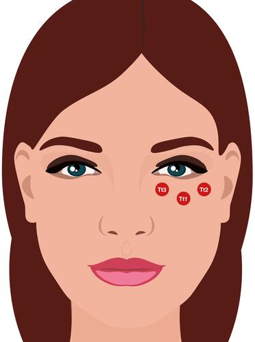
Table 4 Overview of the Different Hyaluronic Acid (HA) Fillers Used in Tear Trough Deformity Treatment and Their Main Characteristics
Table 5 Authors Recommendations for Treating Tear Trough
Table 6 Overview of the Authors’ Recommendations for Treating Tear Trough Deformity with Hyaluronic Acid (HA) Filler According to the MD Codes®.
Figure 4 Overview of the different Codes involved in the tear trough deformity indirect approach treatment. Data from Peng et alCitation6 and de Maio.Citation10 Image courtesy from and reprinted with permission from Allergan plc, Dublin, Ireland.
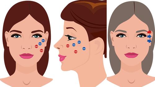
Figure 5 Codes involved in the direct approach strategy for treating tear trough deformity. Tear trough and orbital codes should be reserved for specialists specifically trained in this technique and those who have a sound knowledge of the anatomy and physiology for this particular area. Data from Peng et alCitation6 and de Maio.Citation10 Image courtesy from and reprinted with permission from Allergan plc, Dublin, Ireland. (A) Imaging representing the frontal view. (B) Imaging representing the anatomical structures. Red circle under Tt1 and exclamation mark: Be aware of the infraorbital artery branches. Red circle near to Tt3 and exclamation mark: Be aware of the angular artery and vein. Codes have been adapted from de Maio.Citation10,Citation11
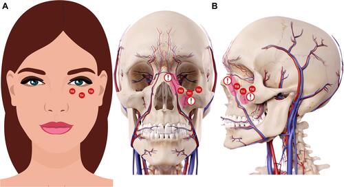
Figure 6 Temporal point to be treated. Data from de Maio.Citation10 Image courtesy from and reprinted with permission from Allergan plc, Dublin, Ireland.

Figure 7 Structural treatment of tear trough deformity. Image courtesy from and reprinted with permission from Allergan plc, Dublin, Ireland. aDo not inject into the cartilage or into the bone, but rather at the level of the cartilage or the level of the bone.
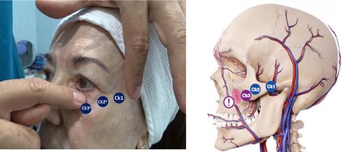
Figure 8 Different anatomical structures and the codes of the periorbital region. Data from de Maio.Citation10 Image courtesy from and reprinted with permission from Allergan plc, Dublin, Ireland.
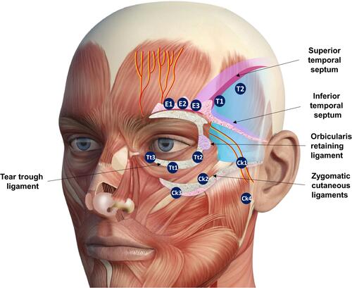
Figure 9 Frontal, right, and left projection of a 40-year-old patient face before (A) and after (B) being treated. The patient provided their consent for the use of their image in this publication. 1. Zygomatic arch (Ck1): 0.3 mL per side of Hyaluronic acid filler (VYC-20L). 2. Anteromedial cheek (Ck3): 0.7 mL per side of Hyaluronic acid filler (VYC-20L). Image courtesy with permission from Dr Farollch. Codes have been adapted from de Maio.Citation10
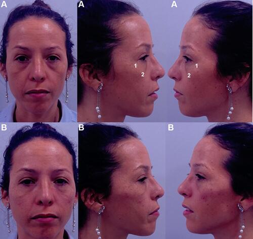
Figure 10 Forty-five years old women with a tear trough deformity (according to Belhaouari classification: 1B (see reference 7)) before treatment (A); that shows the treatment plan (B); and after treatment (C). The patient provided their consent for the use of their image in this publication. 1. Temporo-orbicular cutaneous retaining ligament; 2. Anterior temple (T1); 3. Palpebral ligament; 4. Orbicular retaining ligament; 5. Zygomatic-cutaneous-orbicularis retaining ligament; 6. Zygomatic arch (Ck1); 7. Zygomatic eminence (Ck2); 8. Anteromedial cheek (Ck3); 9. Central infraorbital (Tt1); 10. Medial infraorbital (Tt3). T1 (2): 0.5 mL per side of Hyaluronic acid (HA) filler (VYC-25L; Volux®; Allergan plc, Dublin, Ireland). Ck1 (6): 3 microboluses of 0.1 mL (one in the suture notch and the other two ones to the sides) of HA filler (VYC-20L). Ck2 (7): 0.1 mL of HA filler (VYC-20L). Ck3 (8): 0.2 mL lateral + 0.25 mL medial of HA filler (VYC-20L). Tt1 (9): 0.2 mL of HA filler (VYC-20L). Tt3 (10): 0.2 mL of HA filler (VYC-20L). Image courtesy with permission from courtesy of Dr Urdiales-Gálvez. Codes have been adapted from de Maio.Citation10
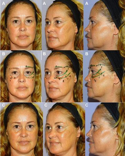
Table 7 Different Aspects That Need to Be Considered When Injecting Hyaluronic Acid (HA) Fillers for Treating Tear Trough Deformity. Adapted from de Maio
Table 8 Overview of the Adverse Events Associated with the Use of Dermal Fillers. Adapted from Funt and Pavicic
Table 9 Overview of the Early and Delayed Adverse Events in Patients Who Underwent Tear Trough Deformity Treatment with Hyaluronic Acid (HA) Fillers