
Figures & data
Figure 1 Case presentation 1. (A) clinical presentation, (B) clinical presentation, detailed view of the suspected lesion, (C) dermoscopic picture of seborrheic keratosis, (D) dermoscopic picture of the pigmented BCC.
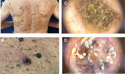
Figure 2 Case presentation 2. (A) clinical presentation, (B) dermoscopic picture of solar lentigo, (C) dermoscopic picture of seborrheic keratosis, (D) dermoscopic picture of the lentigo malignant melanoma.
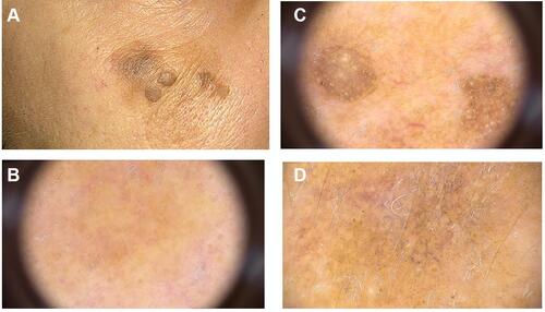
Table 1 Dermoscopic Features of Seborrheic Keratosis
Table 2 Differential Diagnosis of Basal Cell CarcinomaCitation26
Table 3 Dermoscopic Features of Basal Cell Carcinoma and Basal Squamous Cell Carcinoma
Table 4 Clinical Grades and Dermoscopic Patterns of Actinic KeratosisCitation35
Table 5 Differential Diagnosis of Squamous Cell CarcinomaCitation32
Table 6 Dermoscopic Features of Squamous Cell Carcinoma (SCC) and Keratoacanthoma
Table 7 Dermoscopic Features of Melanoma Varieties