Figures & data
Table 1 AOR and Extent of Repigmentation Among Treatment Groups
Figure 1 Changes in the lesional median surface area from baseline to 3 and 6 -months, for the treatment groups.
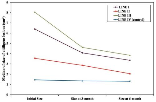
Table 2 Cosmetic Matching Among the Four Lines of Treatment
Figure 2 Vitiligo vulgaris lesion on nape of the neck treated by mini-punch grafts. (A) Baseline surface area of 7.3 cm2. (B): 47.9% Repigmentation after 6 months.
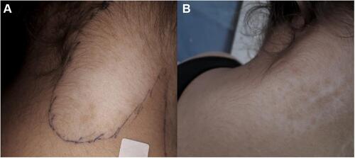
Figure 3 Vitiliginous lesion on left wrist treated with needling (A) initial surface area of 10.2 cm2. (B) 22.5% Repigmentation after 3 months, with partial marginal hyperpigmentation. (C) 51% Repigmentation at 6 months.
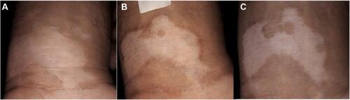
Figure 4 Trunkal vitiligo lesion treated with mini-punch grafts followed by transverse needling sessions. (A) Baseline surface area of 7.9 cm2. (B) 88.6% Repigmentation after 3 months. (C) 91.1% Repigmentation after 6 months with excellent color match.
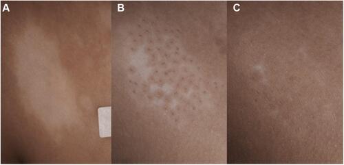
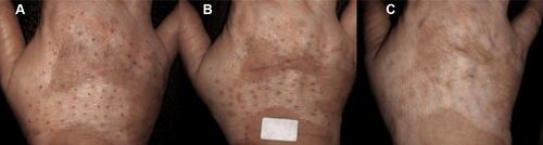
Figure 5 Loss of graft pigment due to lesional activity. (A) Baseline right-hand dorsum vitiligo lesion. (B) Perigraft pigmentation with good marginal definition at 12 weeks after mini-punch grafts followed by needling sessions. (C) Loss of pigmentation at and around the implanted grafts at 18 weeks indicating activity.