Figures & data
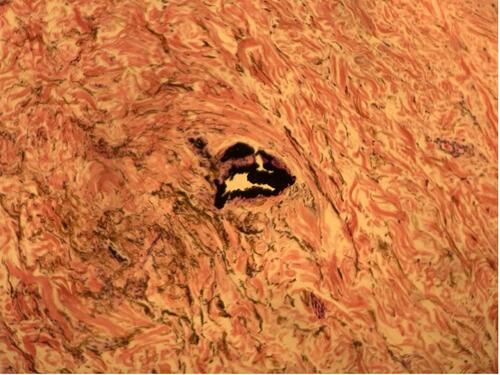
Figure 1 Microscopic appearance of the bluish macule after haematoxylin and eosin staining.
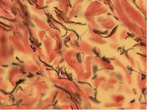
Figure 2 Large, pigmented cluster on the haematoxylin and eosin-stained histological sample.
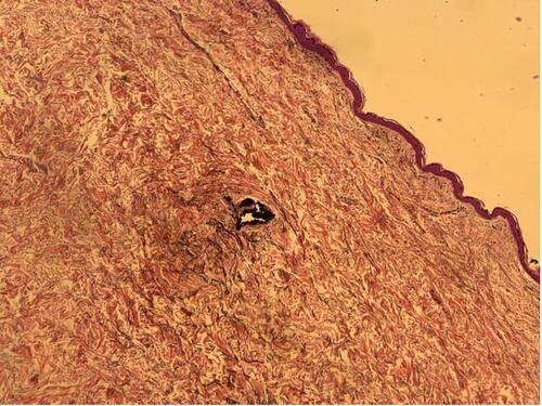
Figure 3 Magnified image of the large, pigmented cluster on the haematoxylin and eosin-stained histological sample.