Figures & data
Figure 1 Expression of melatonin receptor 1A in keratinocytes enriched with Merkel cell culture at passage 1 and 3; DAPI stained nuclei; expression of cytokeratin-20 – green fluorescence; expression of the protein under study – red fluorescence; magnification x40.
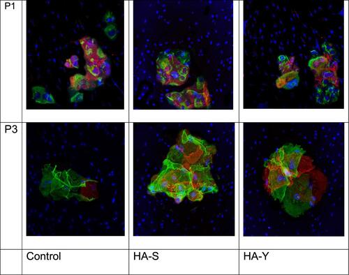
Table 1 The Influence of HA Fillers (HA-Y and HA-S) on the Area of Signaling Molecules Expression in Mixed Culture of Keratinocytes and Fibroblasts
Figure 2 Immunofluorescent analysis of Elastin marker expression in cell culture. Cell nuclei were counterstained with Hoechst. Magnification x40.
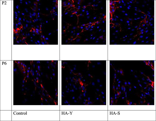
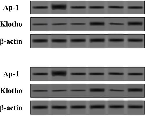
Figure 3 Expression levels of AP-1 and klotho proteins in the mixed culture of human fibroblasts and keratinocytes in the HA-S and HA-Y treated group and the control (Con) group tested by Western blot.