Figures & data
Table 1 The Characteristics of the Participants
Figure 1 (A) Immunohistochemistry (IHC) showing K17 not being expressed in normal healthy skin (NHS); (B) but expressed in hair follicles and sebaceous glands (all magnifications 200×).
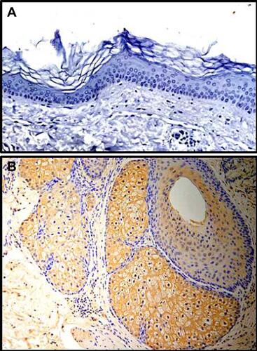
Table 2 The Expression of K17 in the Lesions of CLP, LSC and PN
Table 3 Quantitative Analysis the Expression of K17
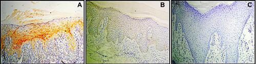
Figure 2 (A) Immunohistochemistry (IHC) demonstrated that K17 was highly expressed in all epidermal layers in cutaneous lichen planus (CLP) lesions (brown staining); (B) but not in lichen simplex chronicus (LSC); (C) and prurigo nodularis (PN). (all magnifications 200×).