Figures & data
Figure 1 Three well-delimited, infiltrated and hard plaques involving the scalp (A), left temple (B) and right temple (C).
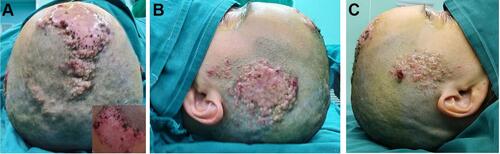
Figure 2 Biopsy, H&E staining, and lamellar bone tissue distributed in the dermis (A). Osteocytes were in the bone tissue, and osteoblasts were seen around bone tissue (B). Black arrows: osteoblast; Green arrows: osteoclast; Red arrow: Lamellar bone tissue.
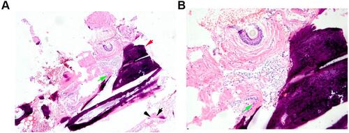
Figure 3 One month after implantation of the dilator. (A) The total amount was 300mL when the water was injected for one month, and the dilator was partially exposed, and the local aseptic bandage was performed. (B) The condition of the skin flap when the expander is removed three months later.
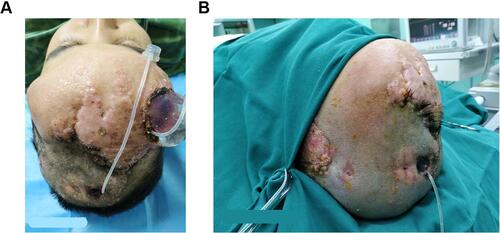
Figure 4 Surgical removal of large skin lesions (A), surgical incisions (B), postoperative sutures (C).
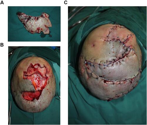
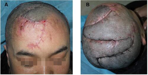
Figure 5 A light red striped scar can be seen on the forehead (A) and top of the head (B) after surgery.