
Figures & data
Figure 1 Clinical presentation: (A) multiple linear and serpiginous purpura on the abdomen, (B) back, (C) left thigh.
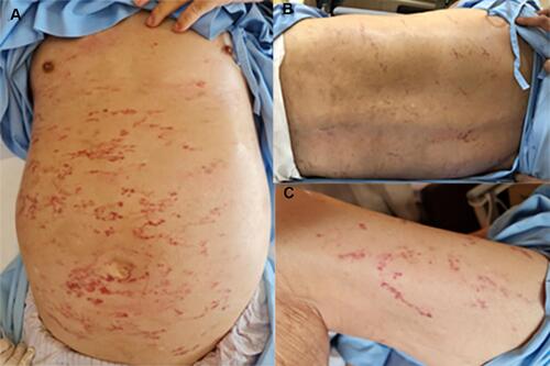
Figure 2 Histopathological and direct immunofluorescence findings: (A) Superficial perivascular infiltration with lymphocytes, neutrophils, nuclear dusts, and extravasated red blood cells, with superficial vascular lumen occlusions. (B) Positive Periodic acid–Schiff (PAS) staining in vessel lumens and vessel walls. (C) Positive IgM staining in vessel lumens and vessel walls. (D) Granular deposition of C3 in superficial vascular walls.
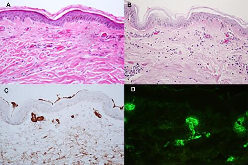
Figure 3 Resolution of lesions on the abdomen.
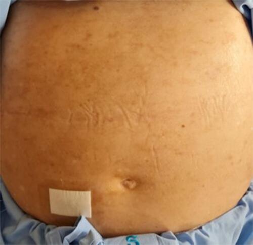
Table 1 Review of Case Reports and Case Series of Cutaneous Macroglobulinosis (CM)