Figures & data
Table 1 Clinical Data and Staining Results of Patients with a Diagnosis of Lentigo Senilis or Lentigo with Mild Junctional Proliferation
Figure 1 Mild atypical junctional proliferation. (A) H&E stain, (B) HMB-45 stain, (C) Melan-A stain, (D) SOX-10 stain. More intense staining noted for Melan-A and SOX-10 at the dermo-epidermal junction. Scale bar = 50 µm.
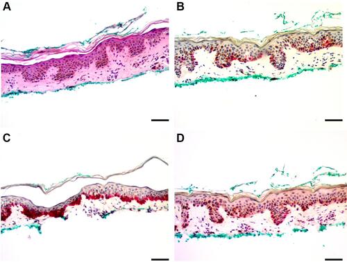
Figure 2 Lentigo with focal junctional melanocytic hyperplasia. (A) H&E stain, (B) HMB-45 stain, (C) Melan-A stain, (D) SOX-10 stain. Melan-A and SOX-10 showed increased staining at the dermo-epidermal junction. Scale bar = 50 µm.
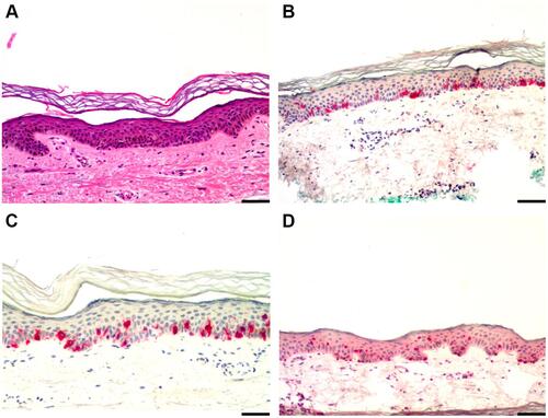
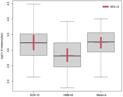
Figure 3 In a logarithmic scale, SOX-10 stained 1.215 times more melanocytes than HMB-45 (p=0.0026) and Melan-A stained 1.214 times more melanocytes than HMB-45 (p=0.0011) with a 95% confidence interval.