Figures & data
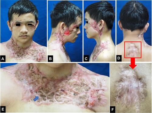
Figure 1 (A–F) Multiple ulcers with granulation tissue on the face, occipital region of the scalp, neck, shoulder, upper chest, and back. (B, C, D and F) A violaceous border appeared around the ulcers (blue arrow). (F) is the enlargement of the region marked with a red square region in (D).
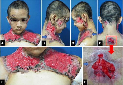
Figure 2 Biopsy specimen from the edge of the ulceration (Hematoxylin and eosin, x10 and x20 magnifications). (A) Histopathological findings showed skin ulceration (asterisk). (B) Massive dermal neutrophilic infiltration mixed with lymphocytic inflammatory infiltrate. (C) Leukocytoclastic vasculitis (blue arrow).
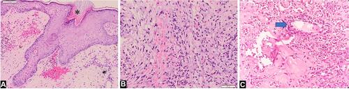
Figure 3 (A–F) After 5 months, cribriform atrophic scars developed at the sites of the previous lesions (blue arrow). (F) is the enlargement of the region marked with a red square in (D).