
Figures & data
Figure 1 Main clinical manifestations. (A and B) Multiple plaques, maculopapules, atrophic scars with bulged edges and partial ulcerated skin lesions on his face; (C and D) Facial skin lesions after 8 sessions of 5-aminolevulinic acid photodynamic therapy.
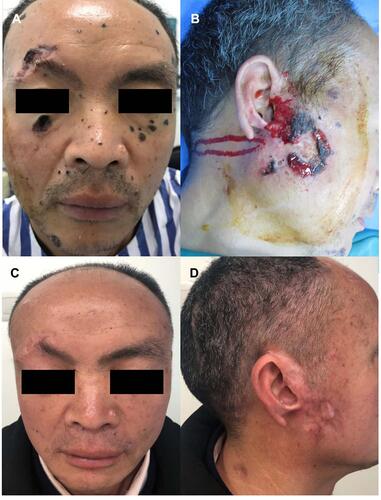
Figure 2 Radiographic findings. (A) Panoramic radiograph of the oral cavity revealed multiple odontogenic keratocysts; (B) Skull computed tomography showed calcification of the falx cerebri; (C–F) Contrast-enhanced computed tomography of the abdomen revealed mesenteric and para-aortic nodules, bilateral renal cysts (the right one-Bosniak grade III).
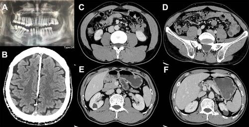
Figure 3 Histopathology of three skin lesions: discrete nests of basaloid cells in the dermis, the peripheral cells are arrayed like a palisade (orange arrows). Adjacent stromal retractions with empty space formations were also noticed (red arrows). Mucin existed among the tumor masses (blue arrow). (hematoxylin eosin, original magnification×40/100). (A–D) face. (E–F) back.
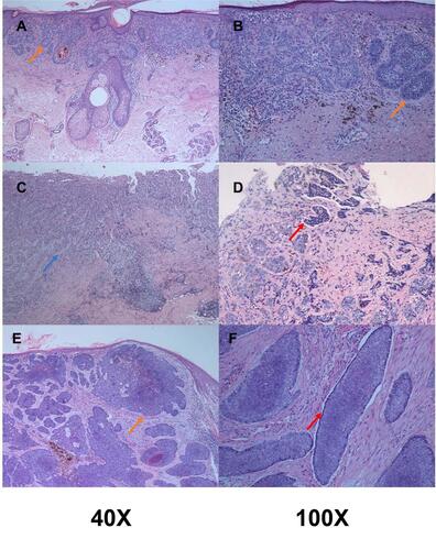
Table 1 Studies of Topical Photodynamic Therapy for Nevoid Basal Cell Carcinoma Syndrome