
Figures & data
Table 1 Cell Differentiation in Dermal Pathology in Melasma
Figure 1 Diagram demonstrated pathologic changes in the dermis of lesional melasma including basement membrane disruption, pendulous melanocytes, melanophages, mast cells, stem cell factor, solar elastosis, and neovascularization.
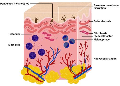
Figure 2 Histopathologic change in the dermis of lesional melasma. (A) Melanin deposition in the epidermis and solar elastosis in the dermis (arrow) (Hematoxylin and Eosin, HEx100) (B) pendulous melanocytes in the basal layer of epidermis (arrow) and increased dermal melanophages (arrowhead) (HE x400).
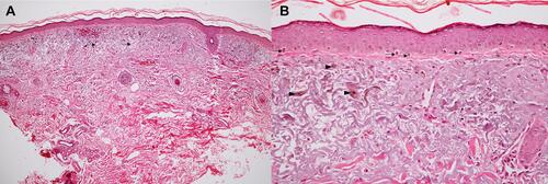
Table 2 The Dermal Pathology in Melasma Studies by the Non-Skin Biopsy Procedure
Table 3 Clinical Application from Dermal Changes in Melasma