Figures & data
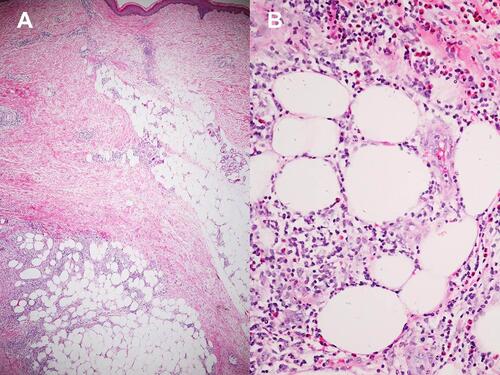
Figure 1 Histopathology of erythema nodosum; (A) Septal panniculitis without vasculitis (Hematoxylin-eosin stain, x40). (B) Higher magnification demonstrating septal fibrosis with lymphohistiocytic infiltration (Hematoxylin-eosin stain, x100).
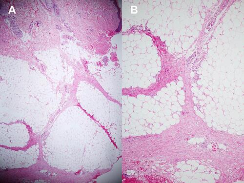
Table 1 Diagnostic Criteria for Diseases and Conditions Associated with Secondary Erythema Nodosum
Figure 2 Recruitment process and the study flow diagram.
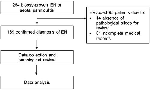
Table 2 Demographic Data, Clinicopathological Results of Erythema Nodosum
Table 3 Etiologies of Erythema Nodosum
Table 4 Treatment of Erythema Nodosum
Table 5 Clinicopathological Findings and Investigations in Relations to Secondary Erythema Nodosum
Figure 3 Erythema nodosum at the dorsum of the left hand in a patient diagnosed with acute myeloid leukemia.
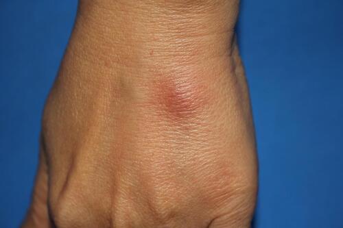
Figure 4 Histopathology of primary erythema nodosum; (A) Septal panniculitis with focal peripheral lobular infiltration (Hematoxylin-eosin stain, x40). (B) Higher magnification demonstrating numerous eosinophils in the area of focal peripheral lobular panniculitis (Hematoxylin-eosin stain, x400).