
Figures & data
Figure 1 Dermatological examination: (A) multiple brown scales with adherent centers and detached, outward-turning edges, with multiple, overlying, small black dots on the extensor areas of both lower extremities; (B) close-up view showing abnormal hairs on the areas of skin abnormality.
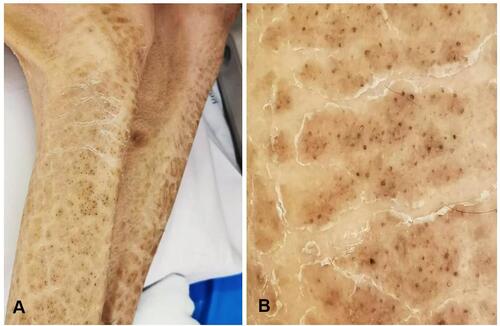
Figure 2 Dermoscopic examination: (A) multiple dark hairs with a perfectly circular arrangement, located under a thin layer of skin (original magnification x20); (B) close-up view of circle hairs (original magnification x80).
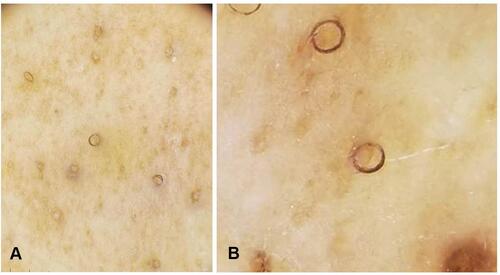
Figure 3 Histopathological examination: laminated hyperkeratosis with hypogranulosis and a dilated hair follicle.
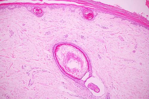
Table 1 Clinical, Dermoscopic, and Histopathologic Features of Circle Hair and Its Mimickers