
Figures & data
Figure 1 Multiple dusky red to brownish papules and patches covered by scales and crusts with some erosions predominately on face (A), upper chest (B), back (C), and dorsum of both forearms (D).
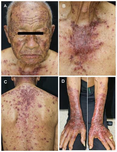
Figure 2 Superficial perivascular infiltration of lymphocytes and melanophages with marked interface change, H&E 100X (A) Epidermal atrophy and few atypical basal keratinocytes, H&E 400X (B).
Abbreviation: H&E, hematoxylin and eosin.
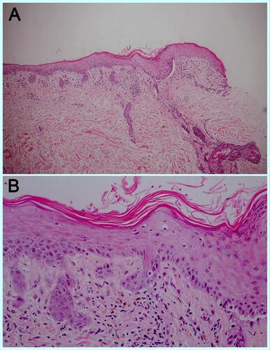
Figure 3 One month after Durvalumab was discontinued, the lesions resolved with post inflammatory hyperpigmentation on face (A), upper chest (B), back (C), and dorsum of both forearms (D).
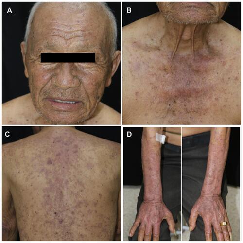
Table 1 Review of Reports on Checkpoint Inhibitors-Associated CLE