Figures & data
Figure 1 The lesion covered with a thick crust (A and B) and ulcer revealed within by removing the crust (C).
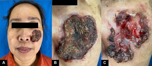
Figure 2 Dermoscopy examination (A) and histopathology examination (B). The dermoscopy, there are a hairpin vessel (black arrow), serpentine vessels (blue arrow), and white structureless area (asterisk).
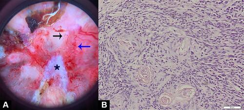
Figure 3 Wide excision and forehead flap. The skin flap was taken from the forehead (A). The skin graft was placed on the forehead to close defect (B).
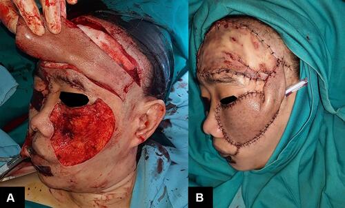
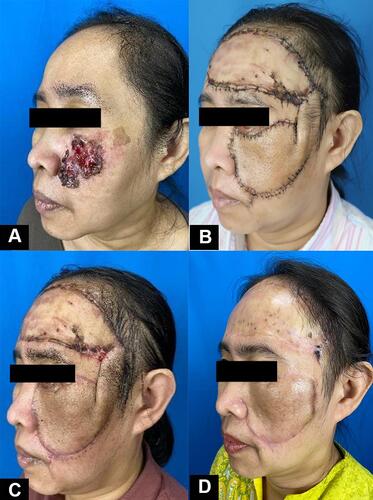
Figure 4 The comparison of the patient before procedure (A), after discharged from inpatient (B), six weeks after procedure (C), and after removal of pedicle (D).